©The Author(s) 2015.
World J Clin Cases. Jun 16, 2015; 3(6): 479-483
Published online Jun 16, 2015. doi: 10.12998/wjcc.v3.i6.479
Published online Jun 16, 2015. doi: 10.12998/wjcc.v3.i6.479
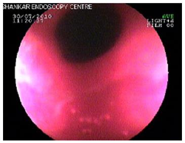
Figure 1 Endoscopic findings include patchy erythematic, gastric outlet narrowing.
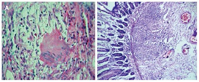
Figure 2 Biopsy showing non-caseating granulomas and oedema in the submucosa (HE × 10).
A: Non-caseating granulomas; B: Oedema alongwith granulation tissue.
- Citation: Ingle SB, Adgaonkar BD, Jamadar NP, Siddiqui S, Hinge CR. Crohn’s disease with gastroduodenal involvement: Diagnostic approach. World J Clin Cases 2015; 3(6): 479-483
- URL: https://www.wjgnet.com/2307-8960/full/v3/i6/479.htm
- DOI: https://dx.doi.org/10.12998/wjcc.v3.i6.479


