©The Author(s) 2015.
World J Clin Cases. Feb 16, 2015; 3(2): 148-155
Published online Feb 16, 2015. doi: 10.12998/wjcc.v3.i2.148
Published online Feb 16, 2015. doi: 10.12998/wjcc.v3.i2.148
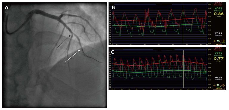
Figure 1 Fractional flow reserve of an “intermediate” lesion in the left anterior descending artery.
A: Fluoroscopic image obtained in the right anterior oblique projection demonstrating an angiographically intermediate stenosis (black arrow) and a pressure wire in-situ (white arrow); B: Pressure trace demonstrating a fractional flow reserve (FFR) of 0.86; C: Pressure trace demonstrating a FFR of 0.77 at maximal hyperaemia that is positive. The patient then proceeded to successful percutaneous coronary intervention of the LAD.
- Citation: Ruparelia N, Kharbanda RK. Role of coronary physiology in the contemporary management of coronary artery disease. World J Clin Cases 2015; 3(2): 148-155
- URL: https://www.wjgnet.com/2307-8960/full/v3/i2/148.htm
- DOI: https://dx.doi.org/10.12998/wjcc.v3.i2.148


