Copyright
©The Author(s) 2015.
World J Clin Cases. Oct 16, 2015; 3(10): 915-919
Published online Oct 16, 2015. doi: 10.12998/wjcc.v3.i10.915
Published online Oct 16, 2015. doi: 10.12998/wjcc.v3.i10.915
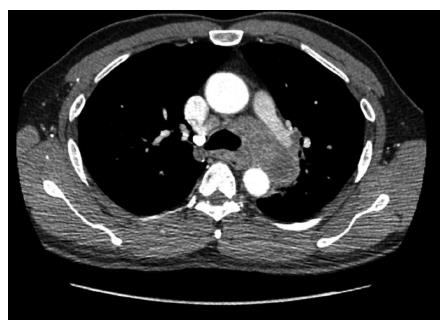
Figure 1 Computed tomography findings.
A 6.6 cm × 4.3 cm mediastinal mass was observed.
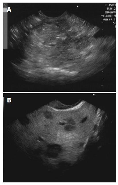
Figure 2 Endoscopic ultrasound findings.
A: Echoview showed a large mediastinal mass; B: Liver nodules with target-like appearance were also observed.
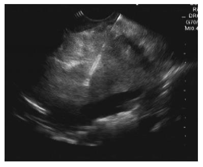
Figure 3 Fine-needle aspiration cytology was performed.
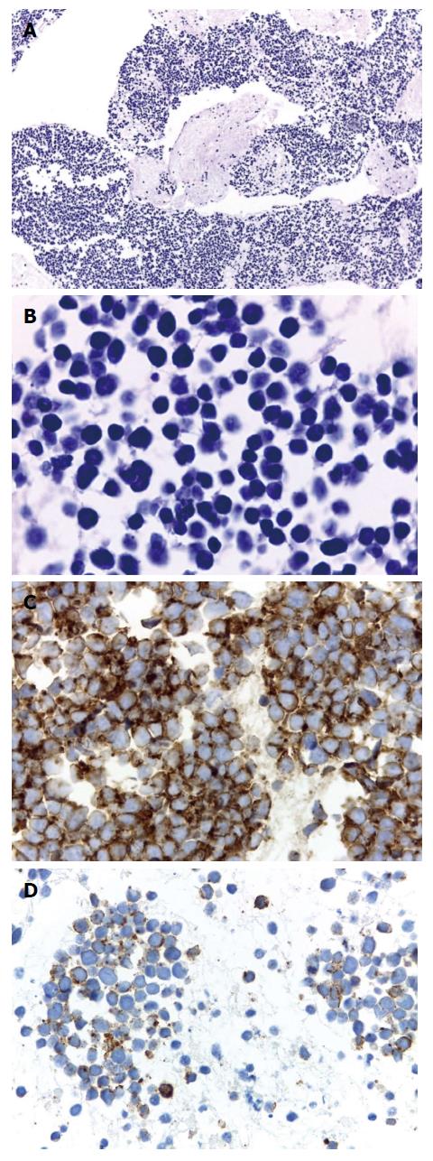
Figure 4 Histopathologic findings.
A and B: Cell-block preparation from fine-needle aspiration of the mediastinal mass showed numerous small, individual, round cells with hyperchromatic nuclei, scarce cytoplasm, and frequent mitotic features. Rare instances of nuclear molding were also observed (hematoxylin-eosin staining, magnification × 10 and × 40, respectively); C and D: Immunohistochemical study revealed that the tumor cells were positive for AE1/AE3 (C) and chromogranin A (D; magnifications × 40).
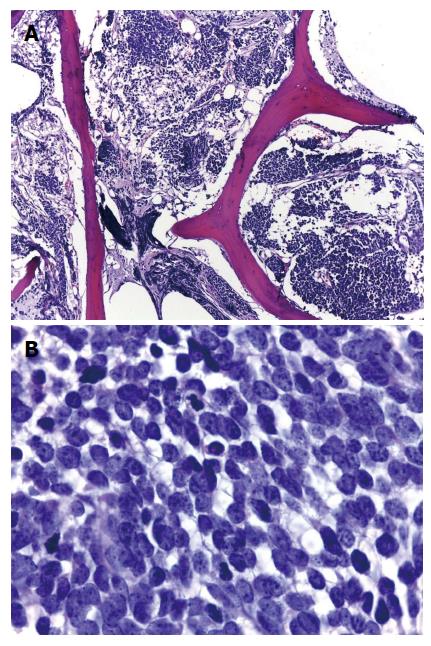
Figure 5 Bone marrow biopsy showed extensive infiltration by cohesive sheets of small, round cells with fine granular chromatin and scarce cytoplasm.
- Citation: Nawarawong N, Pongpruttipan T, Aswakul P, Prachayakul V. Mediastinal small cell carcinoma with liver and bone marrow metastasis, mimicking lymphoma. World J Clin Cases 2015; 3(10): 915-919
- URL: https://www.wjgnet.com/2307-8960/full/v3/i10/915.htm
- DOI: https://dx.doi.org/10.12998/wjcc.v3.i10.915


