©2014 Baishideng Publishing Group Inc.
World J Clin Cases. Aug 16, 2014; 2(8): 391-394
Published online Aug 16, 2014. doi: 10.12998/wjcc.v2.i8.391
Published online Aug 16, 2014. doi: 10.12998/wjcc.v2.i8.391
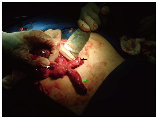
Figure 1 A photograph taken during a laparotomy procedure depicting an inflamed double cecal appendix.
Minor (black arrow) and major (green arrow) inflamed cecal appendix. Surgeon’s hand is on the left side of the picture, holding the proximal segment of the ileum (arrow with white edges).
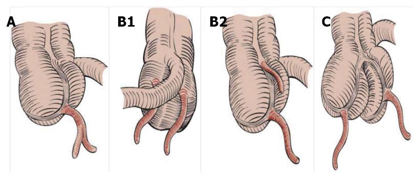
Figure 2 Classification modified by Cave-Wallbridge[30], including type A, subtype B1, subtype B2 and type C.
- Citation: Alves JR, Maranhão &GO, Oliveira PVV. Appendicitis in double cecal appendix: Case report. World J Clin Cases 2014; 2(8): 391-394
- URL: https://www.wjgnet.com/2307-8960/full/v2/i8/391.htm
- DOI: https://dx.doi.org/10.12998/wjcc.v2.i8.391


