
















©2014 Baishideng Publishing Group Inc.
World J Clin Cases. Jul 16, 2014; 2(7): 279-283
Published online Jul 16, 2014. doi: 10.12998/wjcc.v2.i7.279
Published online Jul 16, 2014. doi: 10.12998/wjcc.v2.i7.279
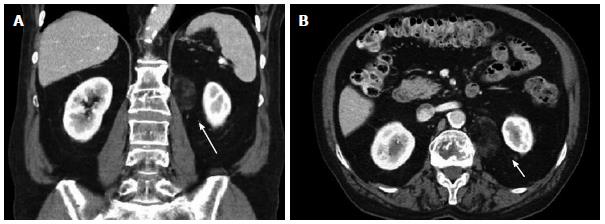
Figure 1 Initial computed tomography scan.
Shows incidentally found non-enhancing heterogeneous mass measuring approximately 3.8 cm × 2.3 cm in longitudinal (A) and anterior-posterior dimensions (B), just inferior to the left renal vein (long arrow) and medial to the left kidney (arrow).
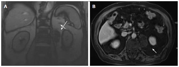
Figure 2 Surveillance magnetic resonance imaging.
Imaging obtained 17 mo after the initial diagnostic computed tomography scan shows that the mass, located just inferior to the left renal vein (long arrow) and medial to the left kidney (arrow), increased in size to 5.0 cm × 3.4 cm in longitudinal (A) and anterior-posterior dimensions (B).
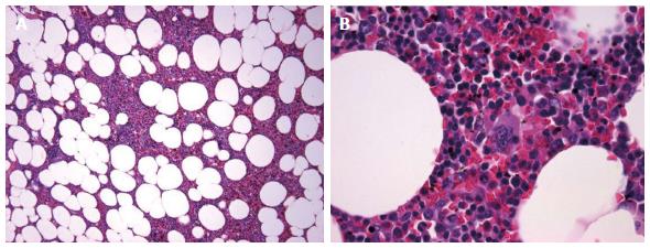
Figure 3 Hematoxylin and eosin stain at 10 × (A) and 60 × (B) magnification.
Shows mature adipose cells with hematopoietic cells including erythroid precursors, granulocytic precursors, and megakaryocytes.
- Citation: Hajiran A, Morley C, Jansen R, Kandzari S, Bacaj P, Zaslau S, Cardinal J. Perirenal extra-adrenal myelolipoma. World J Clin Cases 2014; 2(7): 279-283
- URL: https://www.wjgnet.com/2307-8960/full/v2/i7/279.htm
- DOI: https://dx.doi.org/10.12998/wjcc.v2.i7.279