©2014 Baishideng Publishing Group Co.
World J Clin Cases. Apr 16, 2014; 2(4): 111-119
Published online Apr 16, 2014. doi: 10.12998/wjcc.v2.i4.111
Published online Apr 16, 2014. doi: 10.12998/wjcc.v2.i4.111
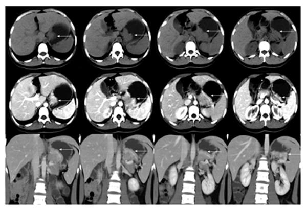
Figure 1 A computed tomography scan showing a mass (5.
8 cm × 3.8 cm × 7.9 cm) in the lesser curvature of the fundus attached to the left adrenal gland with obscure boundaries (white arrows).
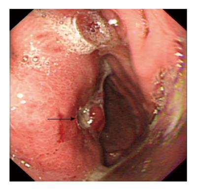
Figure 2 Gastroscopy revealing a protruding lesion on the back wall of the gastric cardia and fundus with a coarse surface, and an approximately 1.
0 cm × 1.0 cm ulcer was located in the middle of the lesion (black arrow).
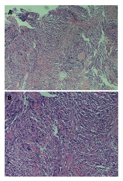
Figure 3 Gastroscopic pathological biopsy findings (Hematoxylin eosin stain) demonstrating an ulcer-forming chronic gastritis without evidence of carcinoma.
[Magnification: A (10 × 10); B (10 × 20)].
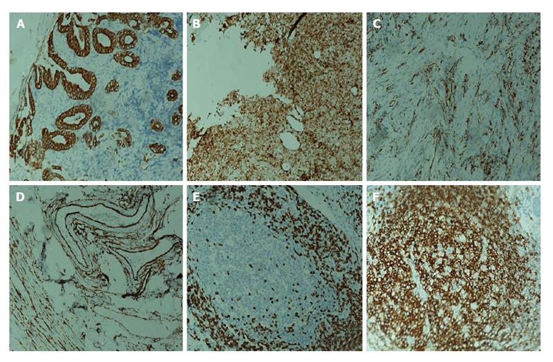
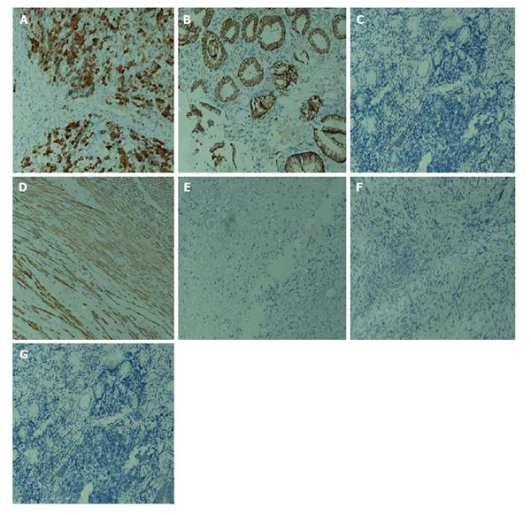
Figure 4 Immunohistochemistry of gastroscopic pathological biopsy.
A: Glandular epithelium CK (+); B: Glandular epithelium M-CEA (+); C: Vascular endothelium CD31 (+); D: Vascular endothelium CD34 (+); E: Small lymphocyte CD3 (+); F: Small lymphocyte L26 (+). Magnification: 10 × 20.
Figure 5 More immunohistochemistry of endoscopic pathological biopsy.
A: Anaplastic lymphoma kinase (+); B: β-catenin (+); C: Desmin (-); D: Actin (+); E: CD117 (-); F: DOG-1 (-); G: S-100 (-). Magnification: 10 × 20.
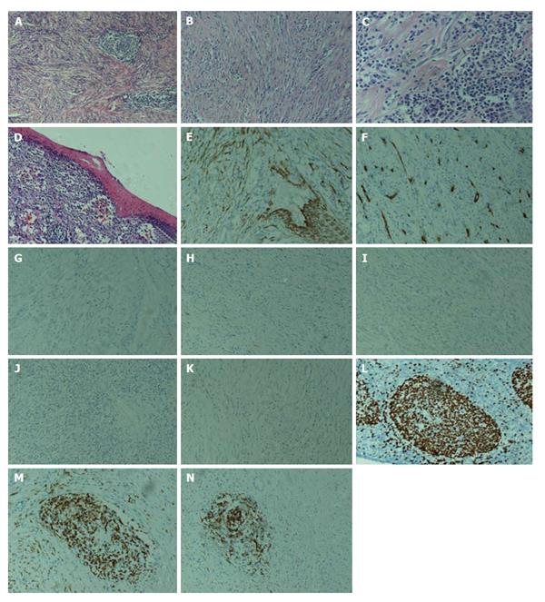
Figure 6 Hematoxylin Eosin stain.
A-D: Microscopically, sub-mucosa composing of mature spindle cells embedded in a dense collagenic hyalinized stroma containing aboundant infiltrative lymphocytes, plasmocytes, and hyperplastic lymphoid follicles with incomplete capsule. No mitosis, necrosis, or nuclear atypia was identified; E-N: Immunohistochemistry, spindle cells: E: Actin focally (+); F: CD34 focally (+); G: CD117 (-); H: S-100 (-); I: desmin (-); J: DOG-1 (-); K: PDGFR (-); L: Ki-67 approximately 2% (+); lymphocytes: M: CD3 (+); N: L26 (+). Magnification: A (10 × 10); B, D-N (10 × 20); C (10 × 40).
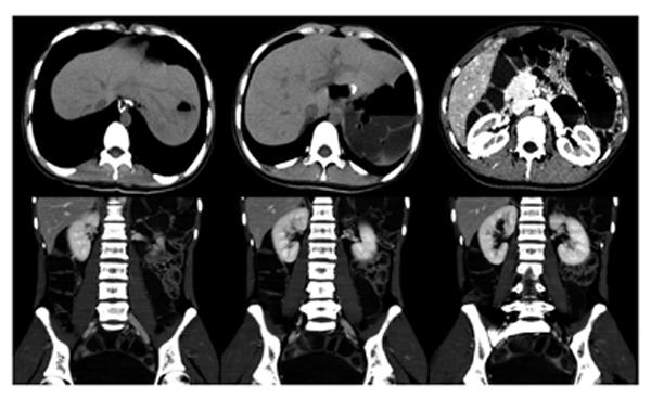
Figure 7 Follow-up abdominal computed tomography scan after 10 mo showing no signs of recurrence and metastatic disease.
- Citation: Yi XJ, Chen CQ, Li Y, Ma JP, Li ZX, Cai SR, He YL. Rare case of an abdominal mass: Reactive nodular fibrous pseudotumor of the stomach encroaching on multiple abdominal organs. World J Clin Cases 2014; 2(4): 111-119
- URL: https://www.wjgnet.com/2307-8960/full/v2/i4/111.htm
- DOI: https://dx.doi.org/10.12998/wjcc.v2.i4.111


