©2014 Baishideng Publishing Group Inc.
World J Clin Cases. Nov 16, 2014; 2(11): 728-731
Published online Nov 16, 2014. doi: 10.12998/wjcc.v2.i11.728
Published online Nov 16, 2014. doi: 10.12998/wjcc.v2.i11.728
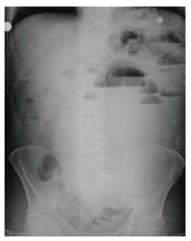
Figure 1 Abdominal radiography, multiple air-fluid levels are seen, which was more prominent in the left upper quadrant.
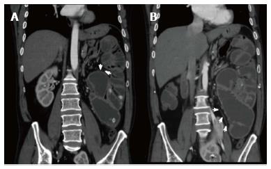
Figure 2 Abdominal computerized tomography - coronal section; dilated small intestinal loops containing air-fluid levels clustered in the left upper quadrant of the abdomen and surrounded by a thick, saclike, contrast-enhanced membrane in the sections close to the root of mesentery (white arrows).
A: Superior; B: Inferior.
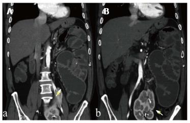
Figure 3 Abdominal computerized tomography - coronal section; dilated small intestinal loops containing air-fluid levels clustered in the left upper quadrant of the abdomen and surrounded by a thick, saclike, contrast-enhanced membrane (A) (arrow).
The left kidney was also located ectopically at the midline in the abdomen at the level of the pelvis (B) (arrow).
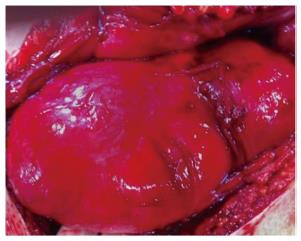
Figure 4 Intraoperative findings; small intestine could not be seen, a capsular structure was identified in the upper left quadrant with a regular surface that was solid-fibrous in nature.
- Citation: Uzunoglu Y, Altintoprak F, Yalkin O, Gunduz Y, Cakmak G, Ozkan OV, Celebi F. Rare etiology of mechanical intestinal obstruction: Abdominal cocoon syndrome. World J Clin Cases 2014; 2(11): 728-731
- URL: https://www.wjgnet.com/2307-8960/full/v2/i11/728.htm
- DOI: https://dx.doi.org/10.12998/wjcc.v2.i11.728


