©2014 Baishideng Publishing Group Inc.
World J Clin Cases. Nov 16, 2014; 2(11): 689-697
Published online Nov 16, 2014. doi: 10.12998/wjcc.v2.i11.689
Published online Nov 16, 2014. doi: 10.12998/wjcc.v2.i11.689
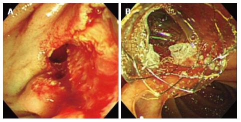
Figure 1 Insertion of fully covered self-expandable metallic stent for the management of periampullary perforation immediately after endoscopic sphincterotomy.
A: A peri-ampullary perforation was seen after endoscopic sphincterotomy; B: A fully covered self-expandable metallic stent (5-cm-long, 10 mm in diameter) inserted into the common bile duct to prevent bile entering the perforation site can be seen at the ampulla of vater.
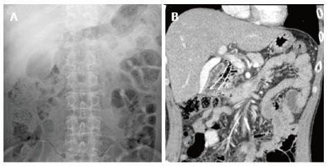
Figure 2 Deployed fully covered self-expandable metallic stent on (A) abdominal X-ray and (B) abdominal computed tomography scan.
- Citation: Lee SM, Cho KB. Value of temporary stents for the management of perivaterian perforation during endoscopic retrograde cholangiopancreatography. World J Clin Cases 2014; 2(11): 689-697
- URL: https://www.wjgnet.com/2307-8960/full/v2/i11/689.htm
- DOI: https://dx.doi.org/10.12998/wjcc.v2.i11.689


