
















©2014 Baishideng Publishing Group Inc.
World J Clin Cases. Nov 16, 2014; 2(11): 683-688
Published online Nov 16, 2014. doi: 10.12998/wjcc.v2.i11.683
Published online Nov 16, 2014. doi: 10.12998/wjcc.v2.i11.683
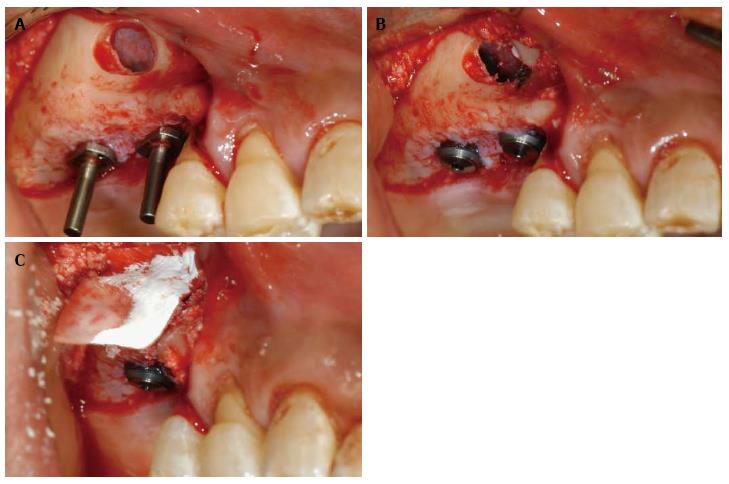
Figure 1 Buccal and clinical view.
A: Buccal view after elevation of sinus membrane; B: Buccal view after installation of dental implants; C: Clinical view after application of graft material.
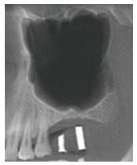
Figure 2 Panoramic view from cone beam computed tomography showing septum.
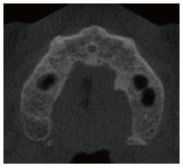
Figure 3 Axial view from cone beam computed tomography with septum on left maxillary sinus.
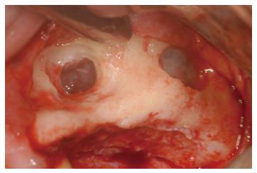
Figure 4 Buccal view showing lateral window design having two windows.
- Citation: Lee JE, Jin SH, Ko Y, Park JB. Evaluation of anatomical considerations in the posterior maxillae for sinus augmentation. World J Clin Cases 2014; 2(11): 683-688
- URL: https://www.wjgnet.com/2307-8960/full/v2/i11/683.htm
- DOI: https://dx.doi.org/10.12998/wjcc.v2.i11.683