©2014 Baishideng Publishing Group Co.
World J Clin Cases. Jan 16, 2014; 2(1): 5-8
Published online Jan 16, 2014. doi: 10.12998/wjcc.v2.i1.5
Published online Jan 16, 2014. doi: 10.12998/wjcc.v2.i1.5
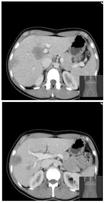
Figure 1 Computed tomography at presentation demonstrating multiple liver lesions.
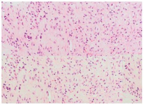
Figure 2 Lesional area composed of cytologically bland, spindle- or stellate-shaped cells arranged in a collagenous stroma with scattered inflammatory cells with predominance of plasma cells.
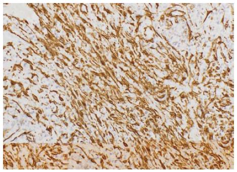
Figure 3 Immunostaining for smooth muscle actin shows that many of the spindled cells are myofibroblasts.
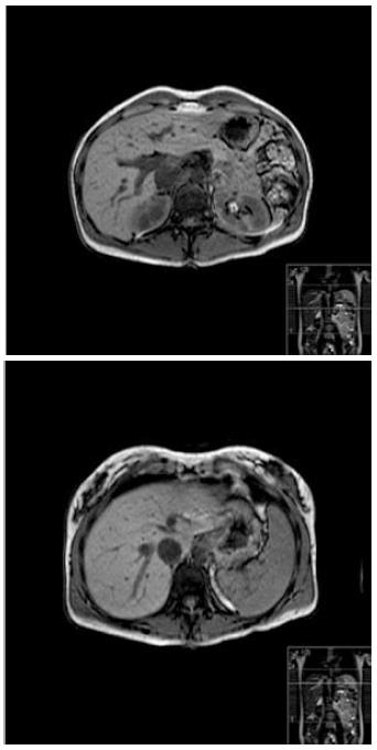
Figure 4 Magnetic resonance imaging of the liver at 2 year follow up demonstrating complete resolution of liver lesions.
- Citation: Puri Y, Lytras D, Luong TV, Fusai GK. Rare presentation of self-resolving multifocal inflammatory pseudo-tumour of liver. World J Clin Cases 2014; 2(1): 5-8
- URL: https://www.wjgnet.com/2307-8960/full/v2/i1/5.htm
- DOI: https://dx.doi.org/10.12998/wjcc.v2.i1.5


