
















Copyright: ©Author(s) 2026.
World J Clin Cases. Oct 26, 2026; 14(28): 127020
Published online Oct 26, 2026. doi: 10.12998/wjcc.127020
Published online Oct 26, 2026. doi: 10.12998/wjcc.127020
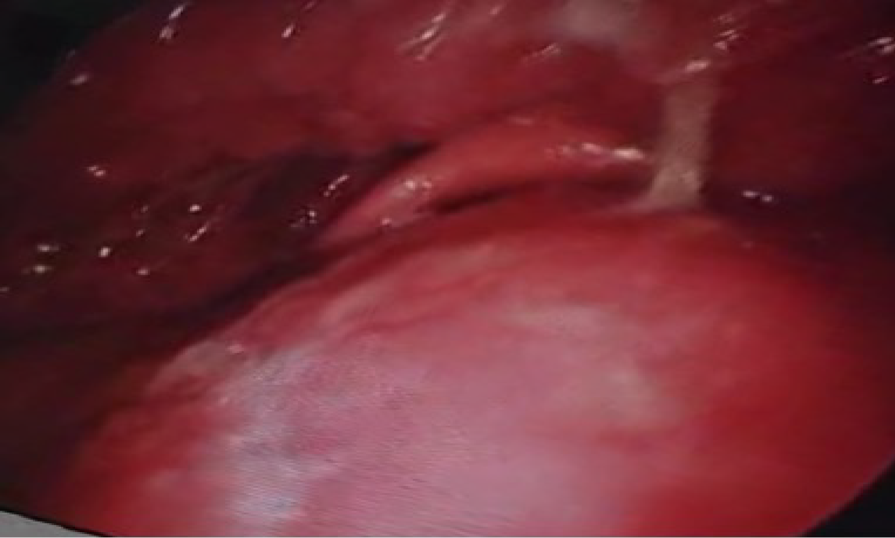
Figure 1
70° endoscopic view of the oropharynx demonstrating a prominent posterior pharyngeal wall bulge with mucosal edema, and non-visualisation of the glottic opening.
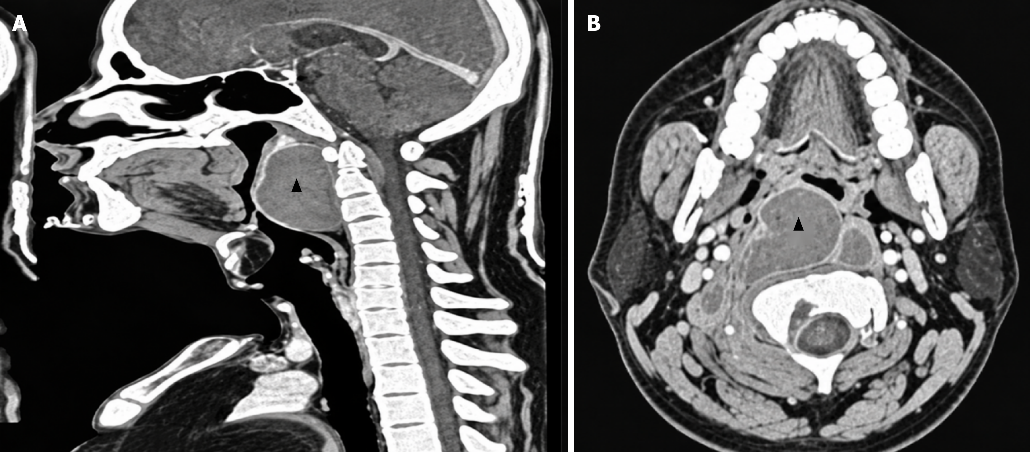
Figure 2 Contrast-enhanced computed tomography of the neck.
A and B: Sagittal (A) and axial (B) images demonstrating a multiloculated parapharyngeal abscess (▲) with extension into the prevertebral and carotid spaces, causing significant narrowing of the oropharyngeal lumen.
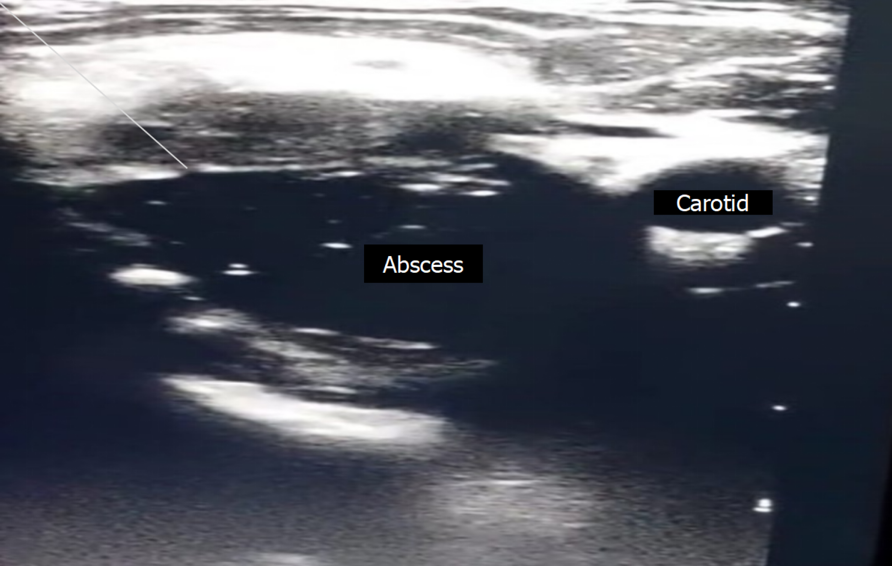
Figure 3
Ultrasound image of the right side of the neck showing a well-defined anechoic collection lateral to the carotid vessels with posterolateral extension, consistent with a parapharyngeal abscess.
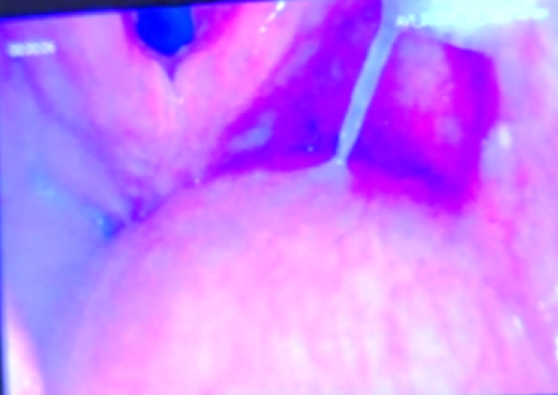
Figure 4
Video-laryngoscopic view obtained after ultrasound-guided aspiration, demonstrating a Cormack-Lehane grade 1 glottic view.
- Citation: Balasubramanian N, Trivedi S, Kushwaha J, Thaware P, Lakra AP, Ankita, Trivedi G. Point-of-care ultrasound-guided aspiration of a parapharyngeal abscess to facilitate airway management: A case report. World J Clin Cases 2026; 14(28): 127020
- URL: https://www.wjgnet.com/2307-8960/full/v14/i28/127020.htm
- DOI: https://dx.doi.org/10.12998/wjcc.127020