
















Copyright: ©Author(s) 2026.
World J Clin Cases. Sep 26, 2026; 14(27): 126689
Published online Sep 26, 2026. doi: 10.12998/wjcc.126689
Published online Sep 26, 2026. doi: 10.12998/wjcc.126689
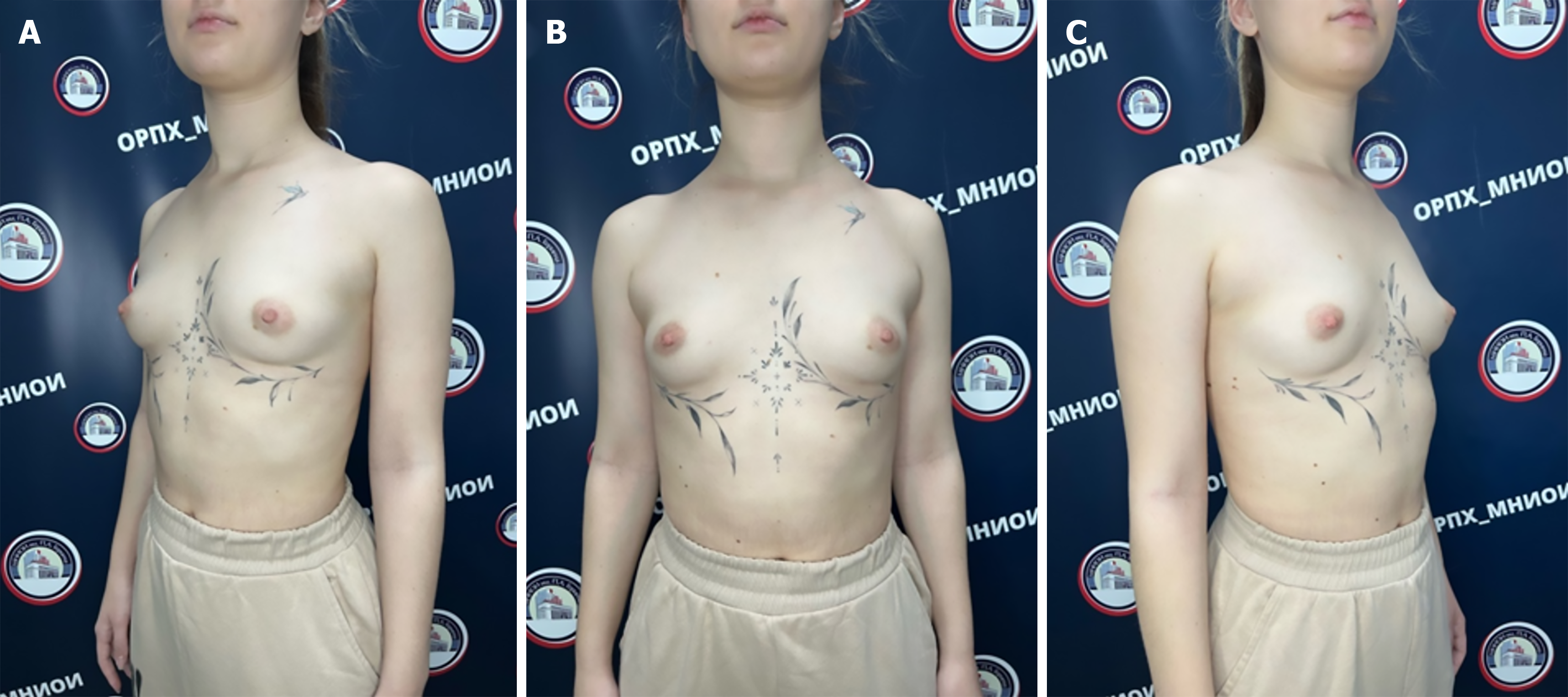
Figure 1 Patient’s appearance before treatment.
A: Anterolateral projection on the left; B: Anterior projection; C: Anterolateral projection on the right.
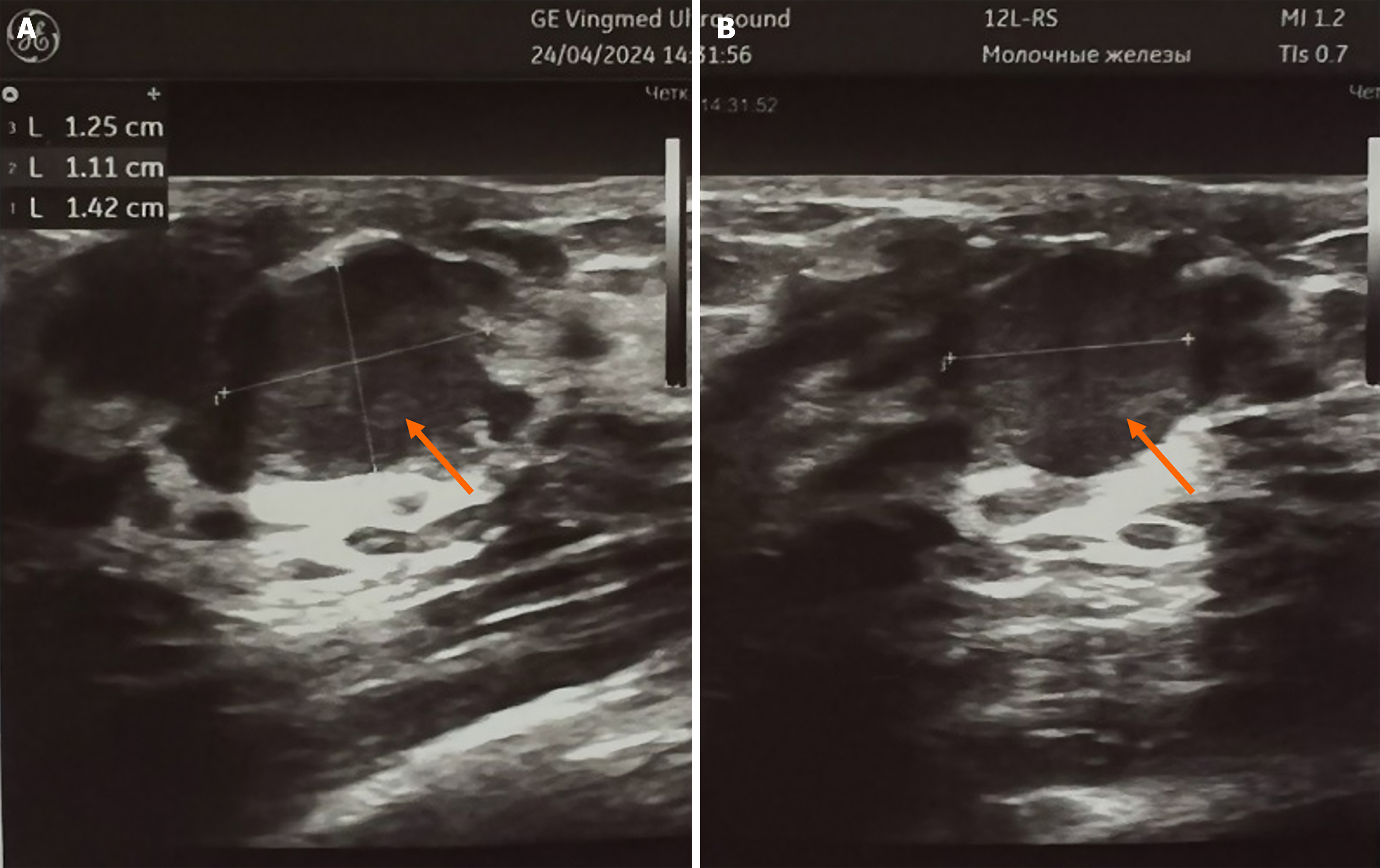
Figure 2 Ultrasound of the mammary glands.
A: Axial position of th probe; B: Saggital position of the probe. A formation located subareolarly in the right mammary gland (indicated by the arrow).
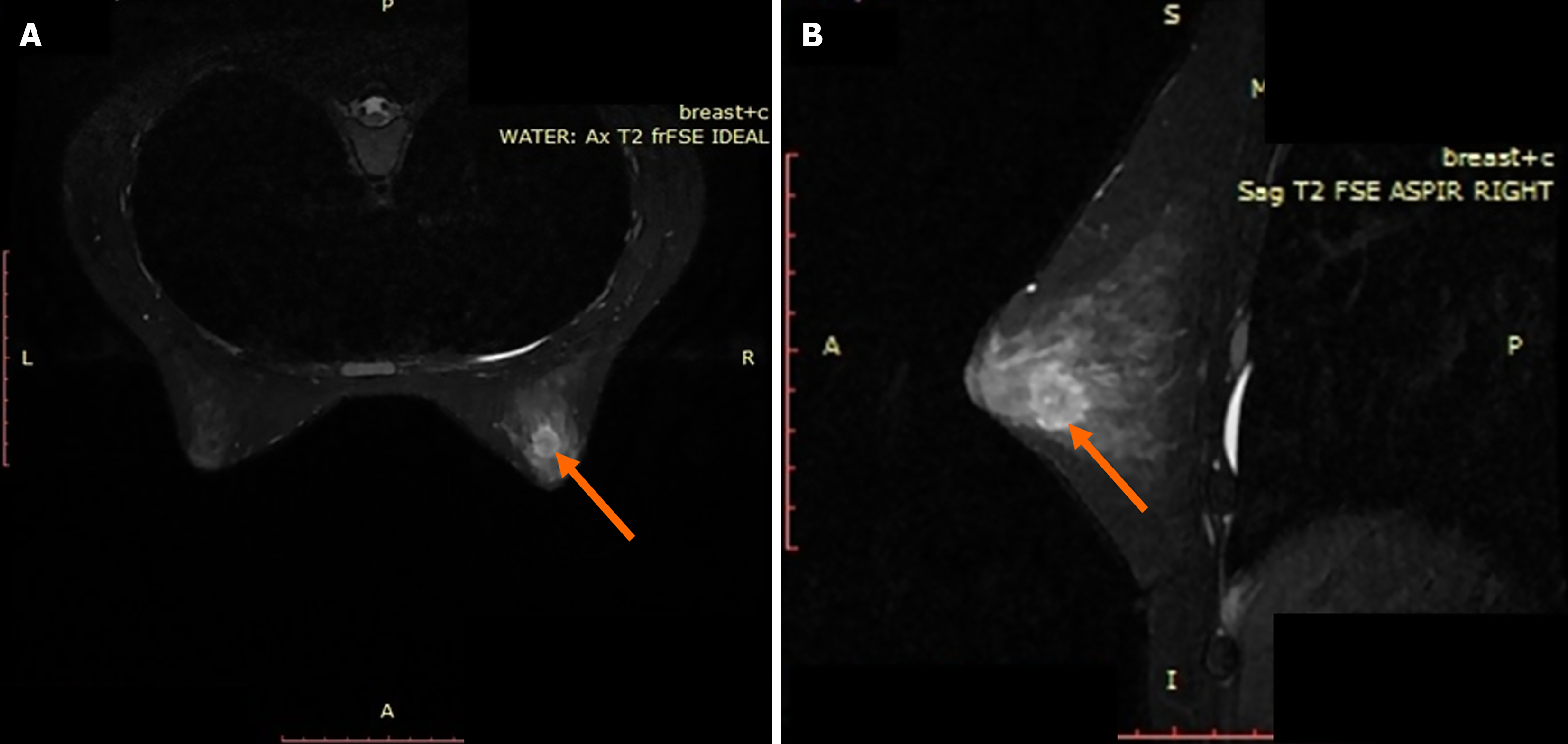
Figure 3 Magnetic resonance imaging of the mammary glands with intravenous contrast enhancement.
A: Direct projection; B: Lateral projection.
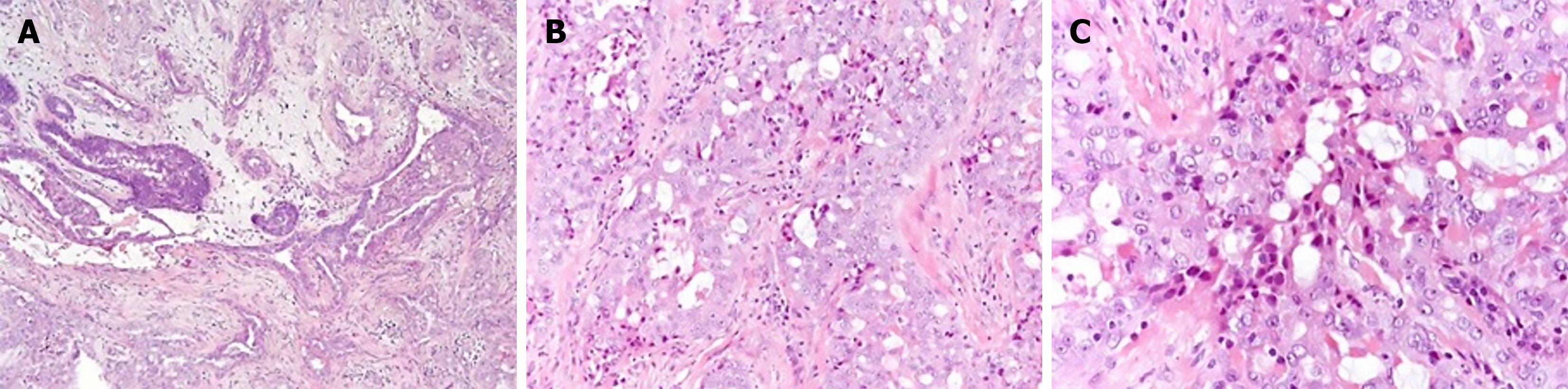
Figure 4 Secretory breast cancer.
A-C: Polygonal cells with eosinophilic granular or vacuolated cytoplasm, round and oval nuclei [A: Hematoxylin and eosin (H&E), ×10; B: H&E, ×20; C: H&E, ×40].
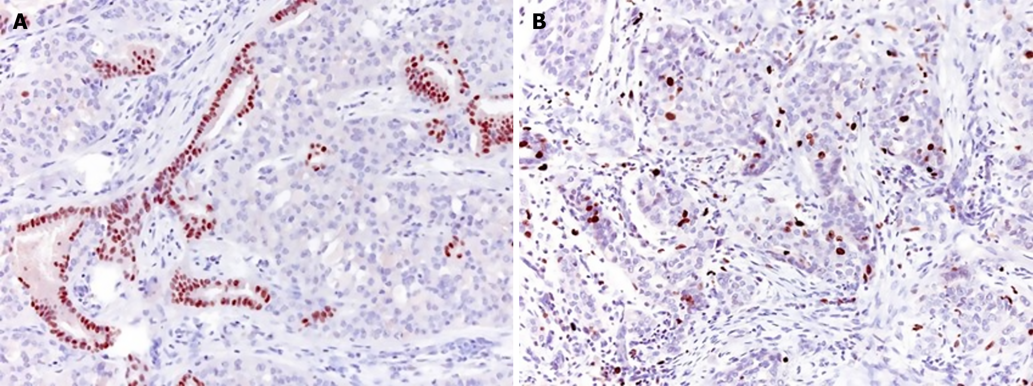
Figure 5 Secretory breast cancer, histological preparations.
A: Positive reaction with estrogen receptors, ×20; B: Expression of Ki 67 in less than 18% of tumor cells, ×20.
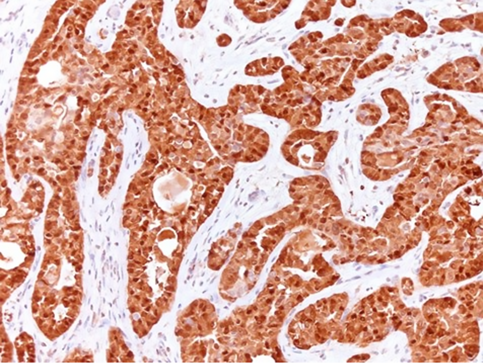
Figure 6 Positive reaction with S 100, ×20.
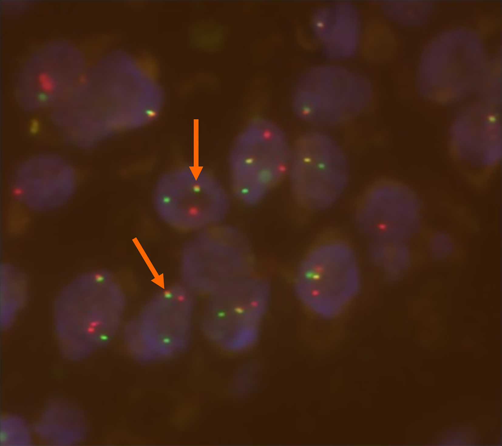
Figure 7 ETV6-NTRK3 gene fusion (marked with arrow).
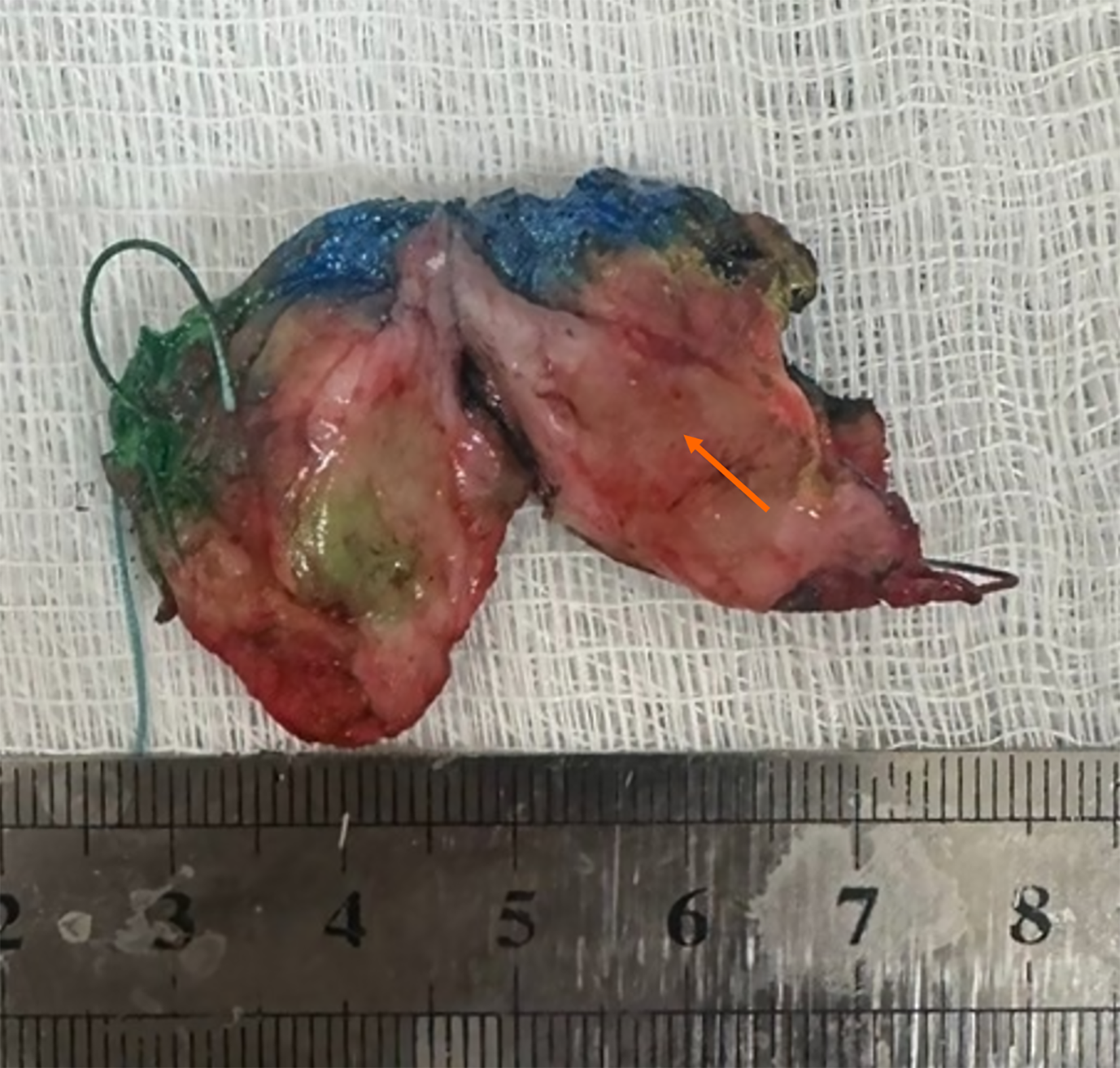
Figure 8 Macroscopic specimen of the removed sector (the tumor node is indicated by an arrow).
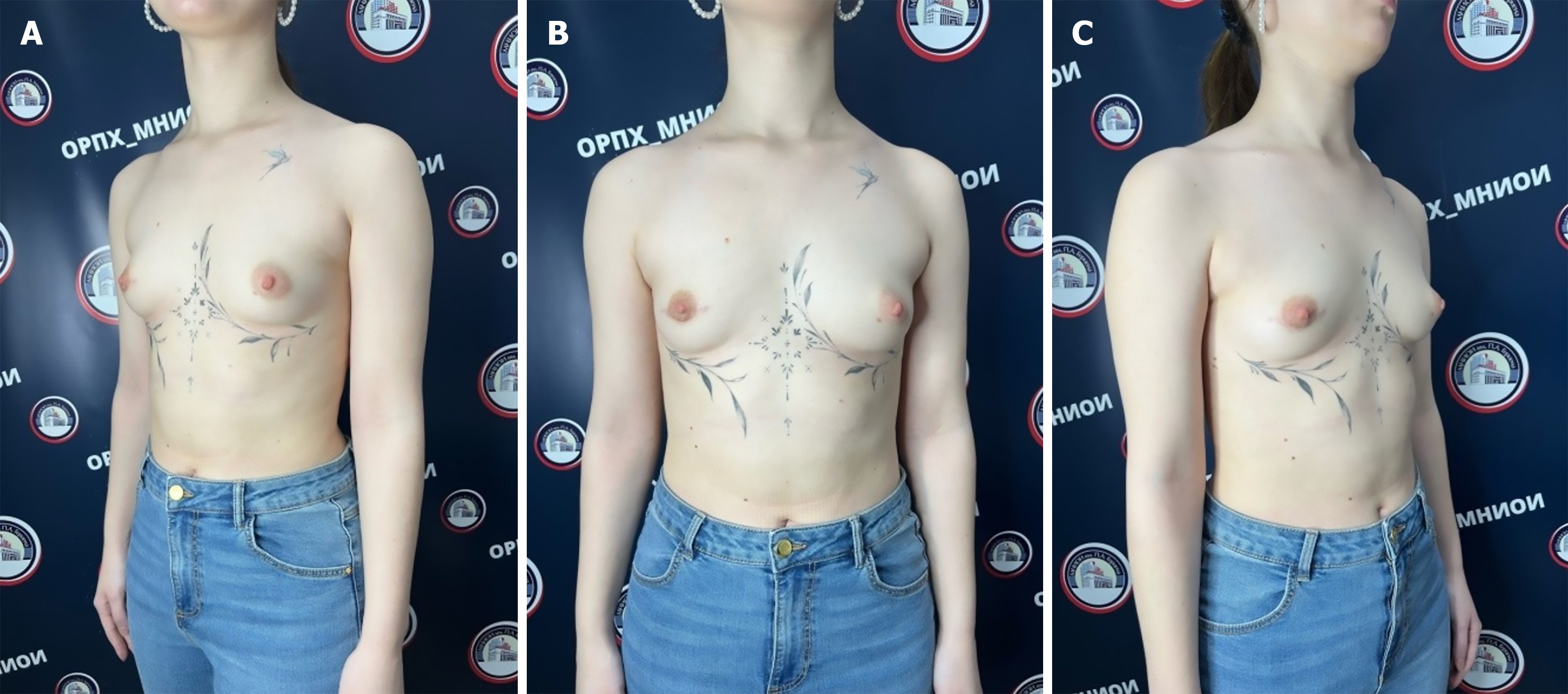
Figure 9 Patient’s appearance 5 months after surgical treatment.
A: Anterolateral projection on the left; B: Anterior projection; C: Anterolateral projection on the right.
- Citation: Naidina K, Ablitsova N, Volchenko N, Goeva N, Razmakhaev G, Zikiryakhodzhaev A, Sukhotko AS, Corlateanu A, Covantsev S. Secretory carcinoma of the breast: A case report. World J Clin Cases 2026; 14(27): 126689
- URL: https://www.wjgnet.com/2307-8960/full/v14/i27/126689.htm
- DOI: https://dx.doi.org/10.12998/wjcc.126689