
















Copyright: ©Author(s) 2026.
World J Clin Cases. Aug 26, 2026; 14(24): 122570
Published online Aug 26, 2026. doi: 10.12998/wjcc.122570
Published online Aug 26, 2026. doi: 10.12998/wjcc.122570
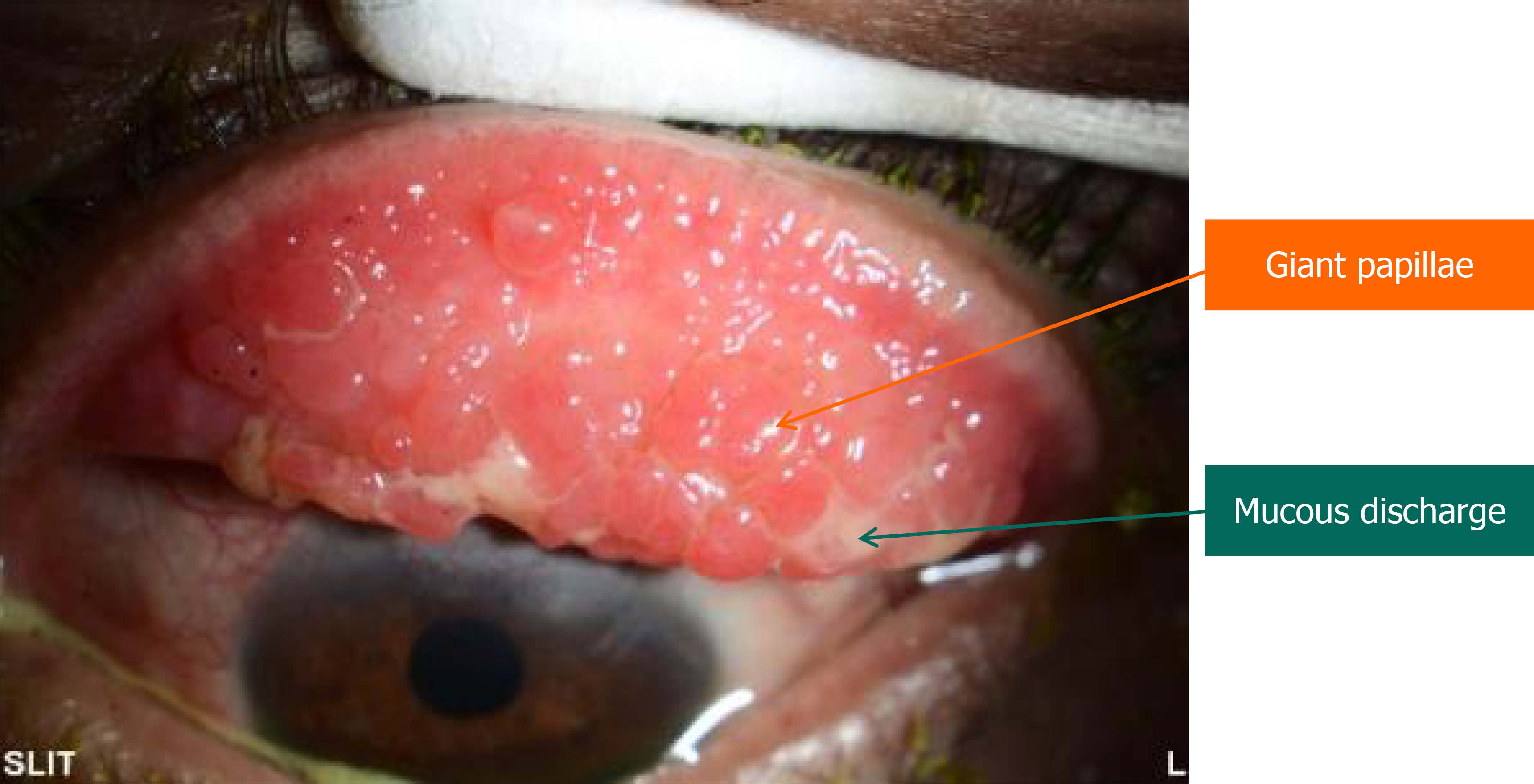
Figure 1
Slit-lamp photograph showing upper lid eversion with multiple giant papillae of the superior tarsal conjunctiva, and mucous discharge in the left eye.
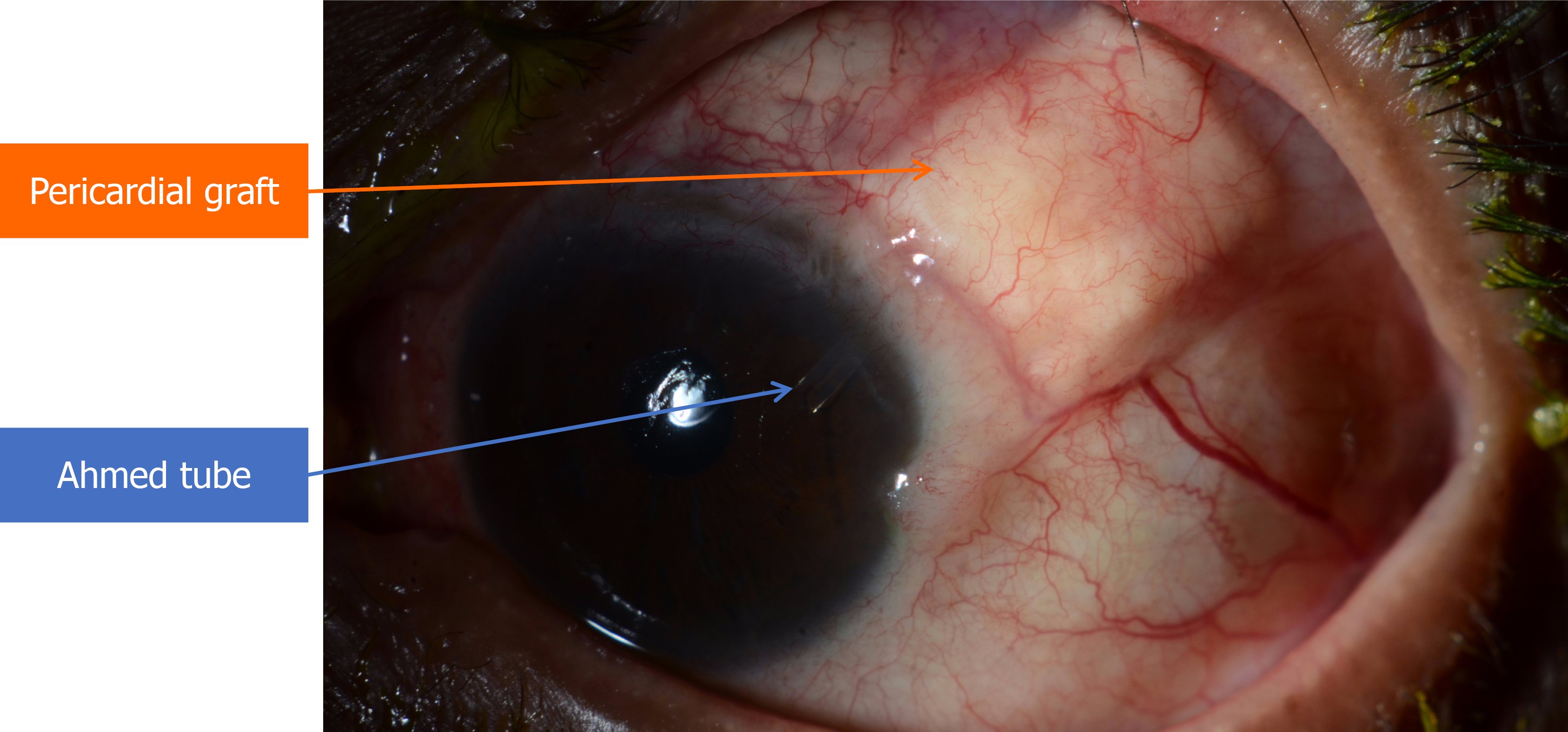
Figure 2
Slit-lamp photograph showing the Ahmed tube in the anterior chamber (blue arrow) and intact double-layer processed pericardial patch graft coverage (orange arrow) with no evidence of exposure.
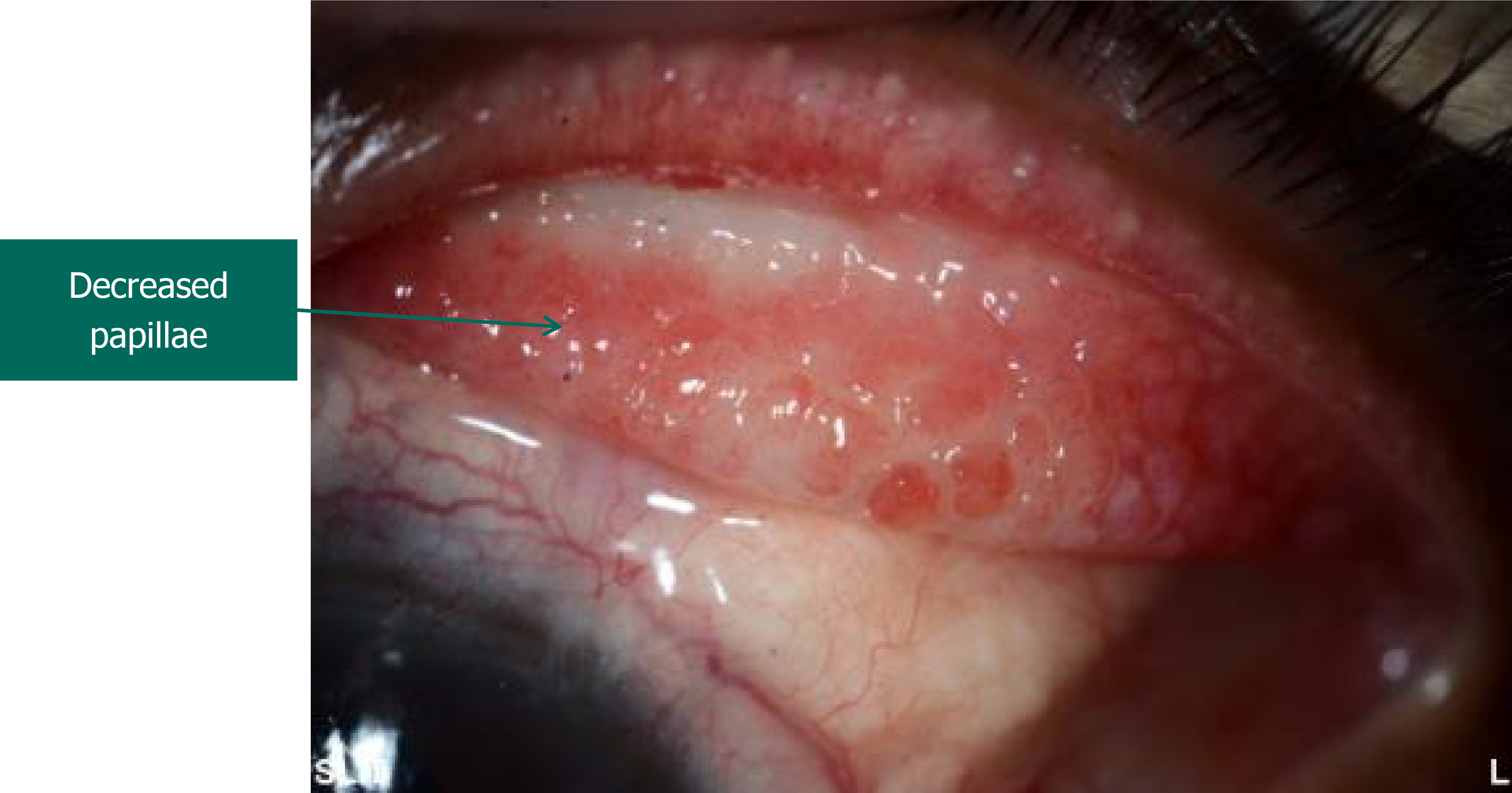
Figure 3
Slit-lamp photograph of the same eye two weeks after initiation of treatment, demonstrating decrease of conjunctival papillae size.
- Citation: Alsirhy E, Alromaih A, Almutairi F, Altoub H. Giant papillary conjunctivitis after Ahmed glaucoma valve implantation with double-layer pericardial patch graft tube coverage: A case report. World J Clin Cases 2026; 14(24): 122570
- URL: https://www.wjgnet.com/2307-8960/full/v14/i24/122570.htm
- DOI: https://dx.doi.org/10.12998/wjcc.122570