
















Copyright: ©Author(s) 2026.
World J Clin Cases. Aug 26, 2026; 14(24): 120680
Published online Aug 26, 2026. doi: 10.12998/wjcc.120680
Published online Aug 26, 2026. doi: 10.12998/wjcc.120680
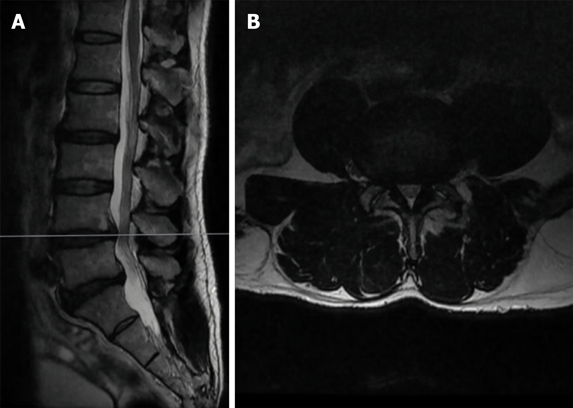
Figure 1 Preoperative magnetic resonance imaging demonstrating bilateral subarticular recess and central canal stenosis.
A: Sagittal view; B: Axial view.
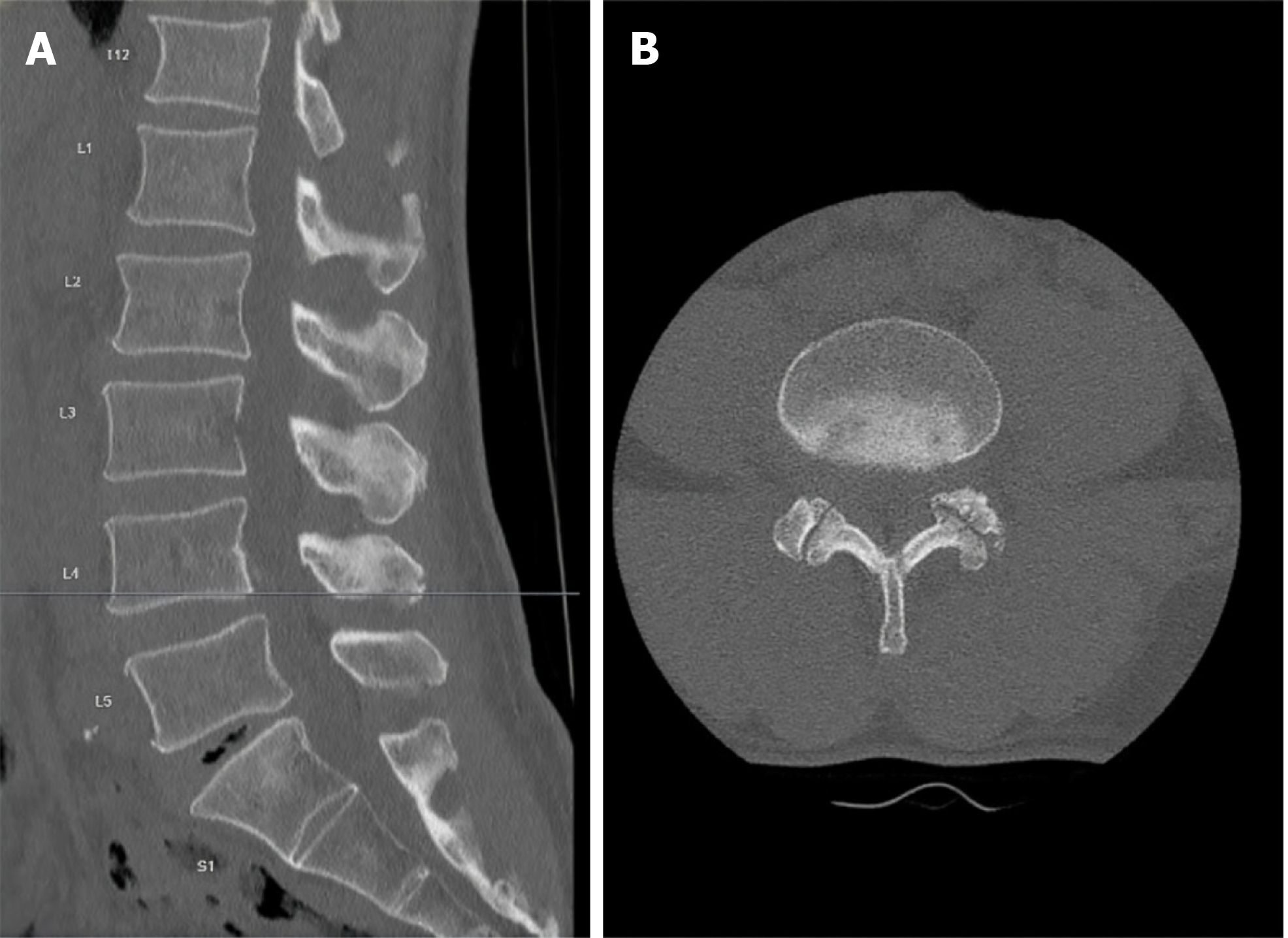
Figure 2 Preoperative thin-cut computed tomography.
A: Sagittal view; B: Axial view.
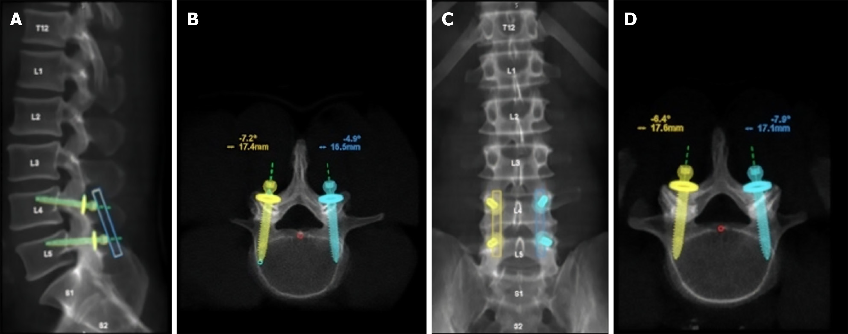
Figure 3 Preoperative hardware planning.
A: Sagittal trajectory; B: Axial trajectory at L4; C: Anteroposterior view; D: Axial trajectory at L5.
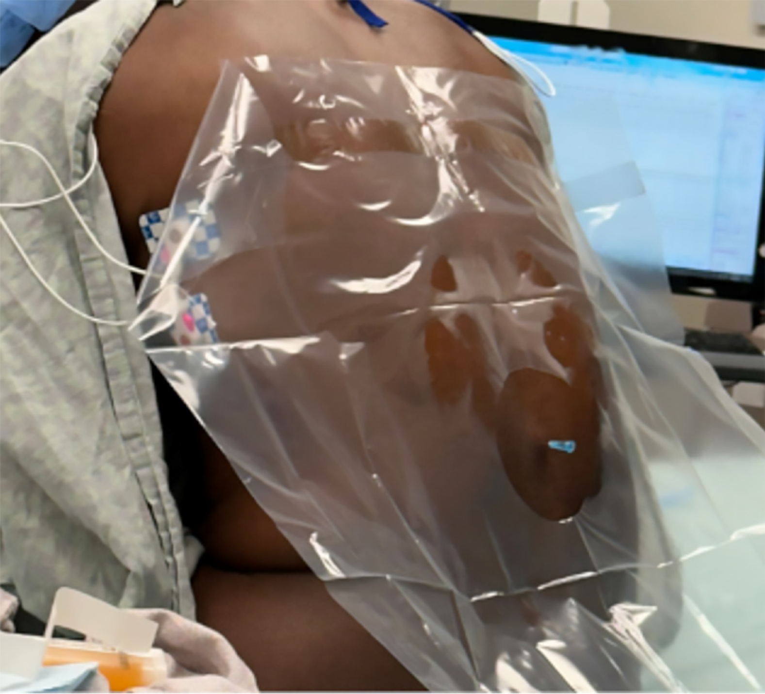
Figure 4 Spinal anesthesia in the sitting position with the spinal needle at the L3-L4 level.
The patient monitor in the background displays real-time vital signs, illustrating active awake intraoperative monitoring.
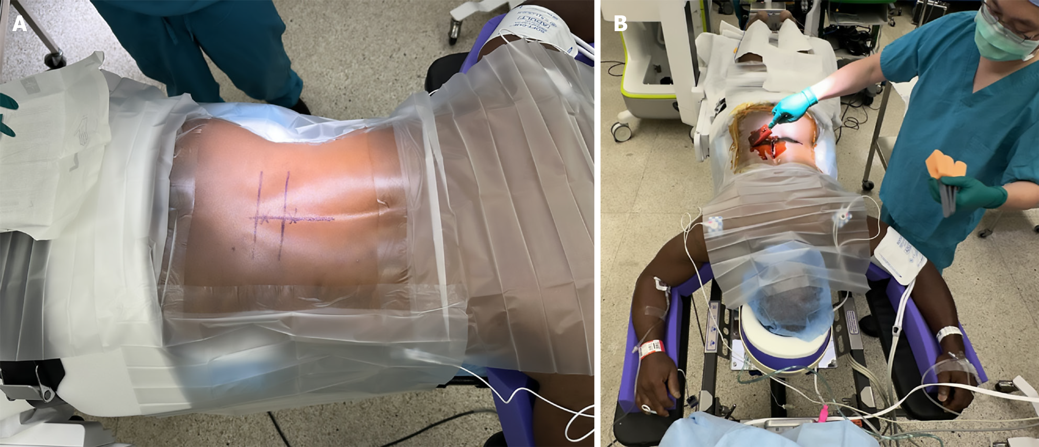
Figure 5 Patient preparation.
A: Skin marking; B: Patient positioning and surgical site preparation.
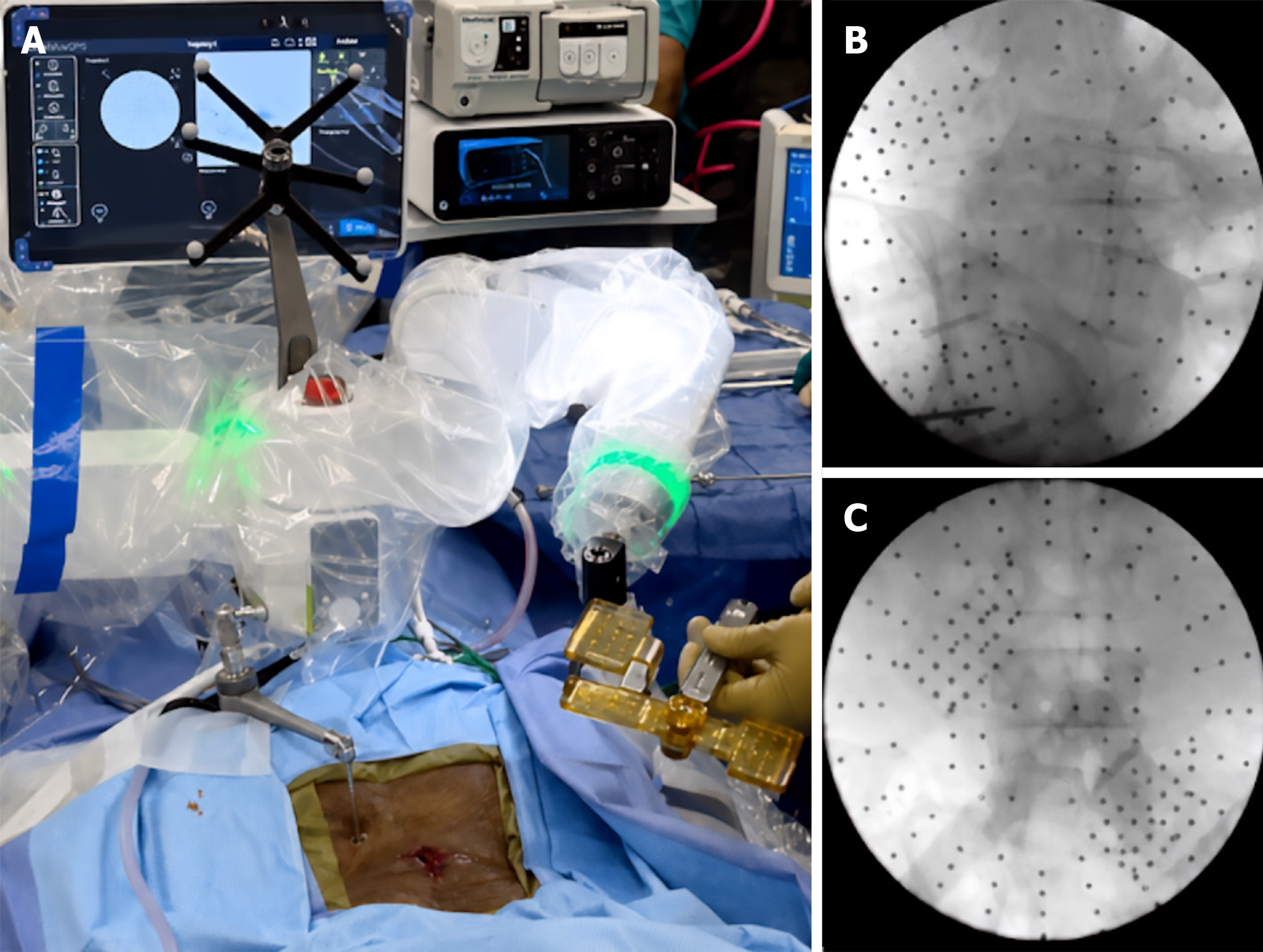
Figure 6 Intraoperative registration.
A: Three-dimensional marker; B: Lateral spinal-level confirmation; C: Anteroposterior spinal-level confirmation.
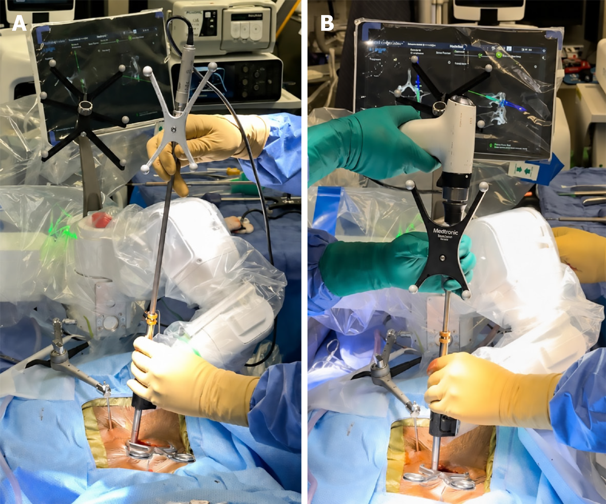
Figure 7 Surgical hardware placement.
A: Drill guide; B: Cortical screw tapping.
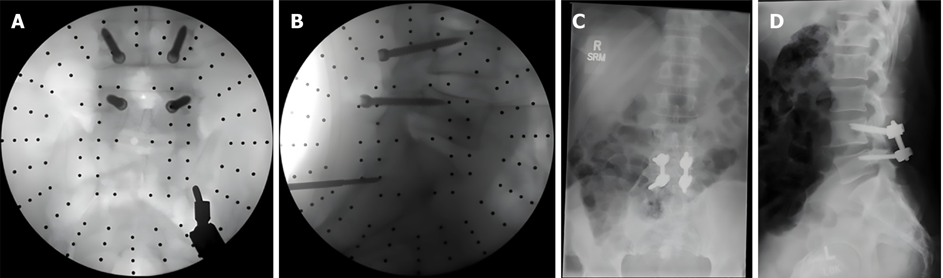
Figure 8 Hardware placement radiographs.
A: Intraoperative anteroposterior view; B: Intraoperative lateral view; C: Postoperative anteroposterior view; D: Postoperative lateral view.
- Citation: Daigle A, Quinones C, Tran H, Whipple G, Kumbhare D, Allampalli V, Guthikonda B, Hoang S. Awake robotic lumbar decompression and fusion: A case report. World J Clin Cases 2026; 14(24): 120680
- URL: https://www.wjgnet.com/2307-8960/full/v14/i24/120680.htm
- DOI: https://dx.doi.org/10.12998/wjcc.120680