
















Copyright: ©Author(s) 2026.
World J Clin Cases. Aug 16, 2026; 14(23): 121732
Published online Aug 16, 2026. doi: 10.12998/wjcc.121732
Published online Aug 16, 2026. doi: 10.12998/wjcc.121732
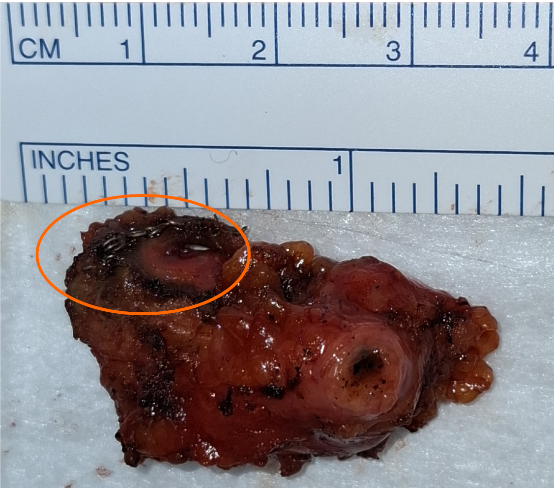
Figure 1 Gross specimen of the gallbladder remnant cystic duct stump following completion cholecystectomy.
The orange circle indicates the metallic clips from the previous subtotal cholecystectomy.
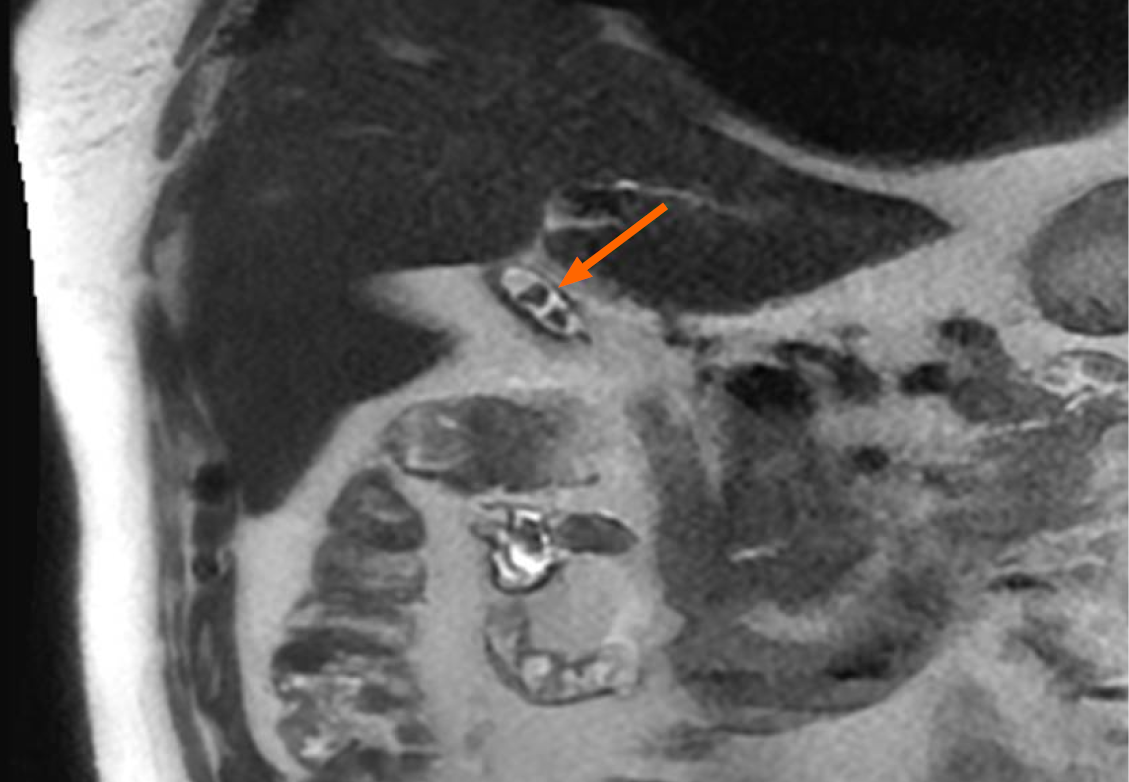
Figure 2 Preoperative magnetic resonance cholangiopancreatography demonstrating an anomalous cystic duct with microlithiasis.
The orange arrow indicates the gallbladder remnant with stones.
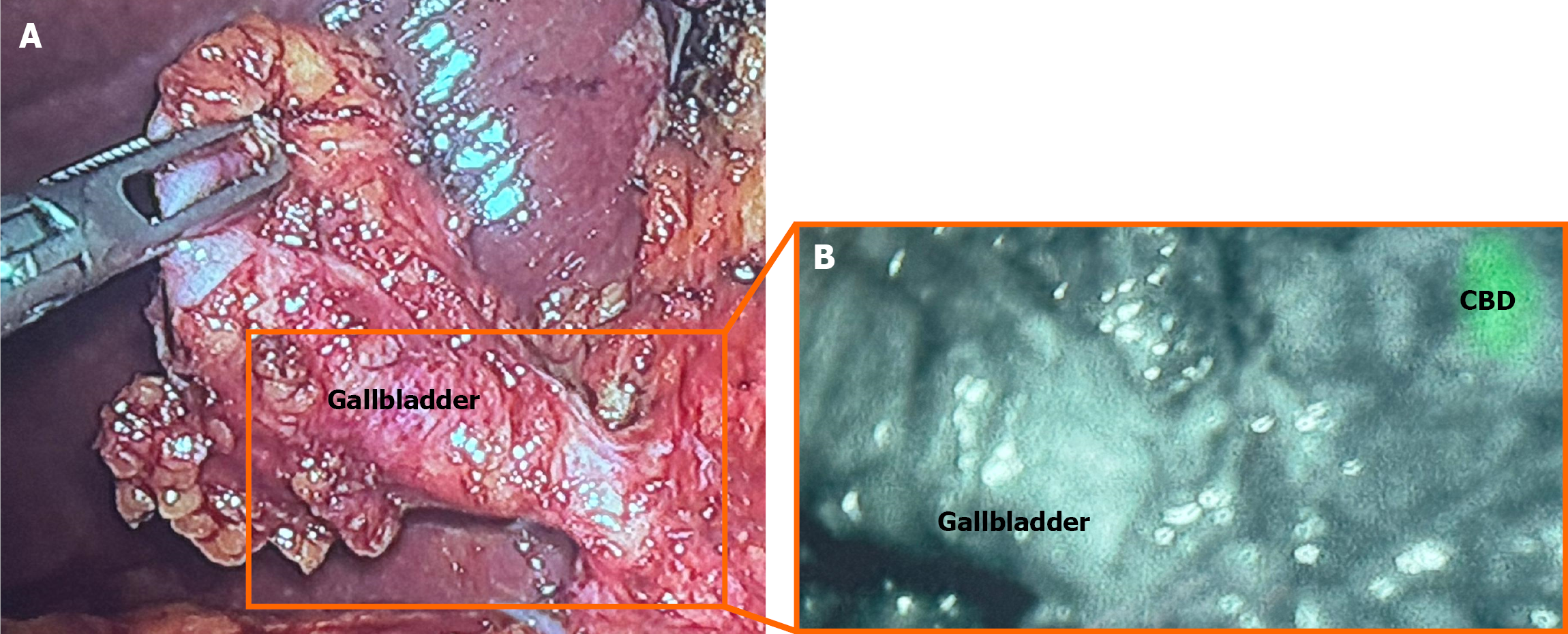
Figure 3 Laparoscopic completion cholecystectomy with indocyanine green fluorescence guidance.
A: Conventional laparoscopic view of Calot’s triangle during reoperation; B: Indocyanine green fluorescence image showing the relationship between the gallbladder remnant and the common bile duct. CBD: Common bile duct.
- Citation: Benea I, Fabbri N, Pesce A, Feo CV. Indocyanine green-guided laparoscopic completion cholecystectomy for stump syndrome: A case report. World J Clin Cases 2026; 14(23): 121732
- URL: https://www.wjgnet.com/2307-8960/full/v14/i23/121732.htm
- DOI: https://dx.doi.org/10.12998/wjcc.121732