
















Copyright: ©Author(s) 2026.
World J Clin Cases. Aug 6, 2026; 14(22): 122012
Published online Aug 6, 2026. doi: 10.12998/wjcc.122012
Published online Aug 6, 2026. doi: 10.12998/wjcc.122012
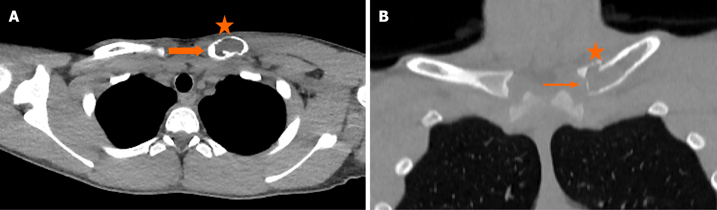
Figure 1 Axial computed tomography and coronal computed tomography chest.
A: Axial computed tomography (CT) chest shows a lytic lesion (asterisk, 4 cm × 2 cm) that affects the medial aspect of the left clavicle with cortical diaphyseal expansion (thick arrow) reaching the inner surface of the sternoclavicular joint; B: Coronal CT chest shows perilesional cortical thinning (asterisk) with focal cortical disruption (slim arrow) and mild periosteal reaction.
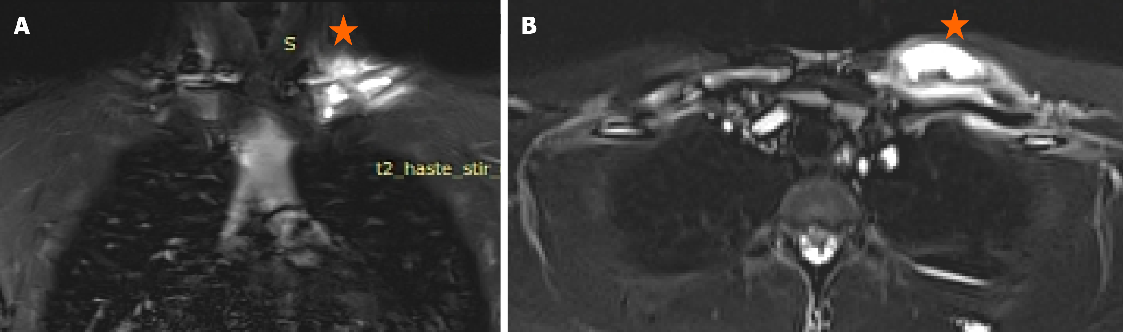
Figure 2 Magnetic resonance imaging at the level of the clavicle.
A and B: Coronal synth short-tau inversion recovery (STIR) (A) and axial STIR (B) images show an intramedullary lesion (asterisk) with abnormal high signal (asterisk) in the anatomical site of the medial aspect of the left clavicle with marked reaction from the surrounding periosteum and adjacent muscles without soft tissue component.
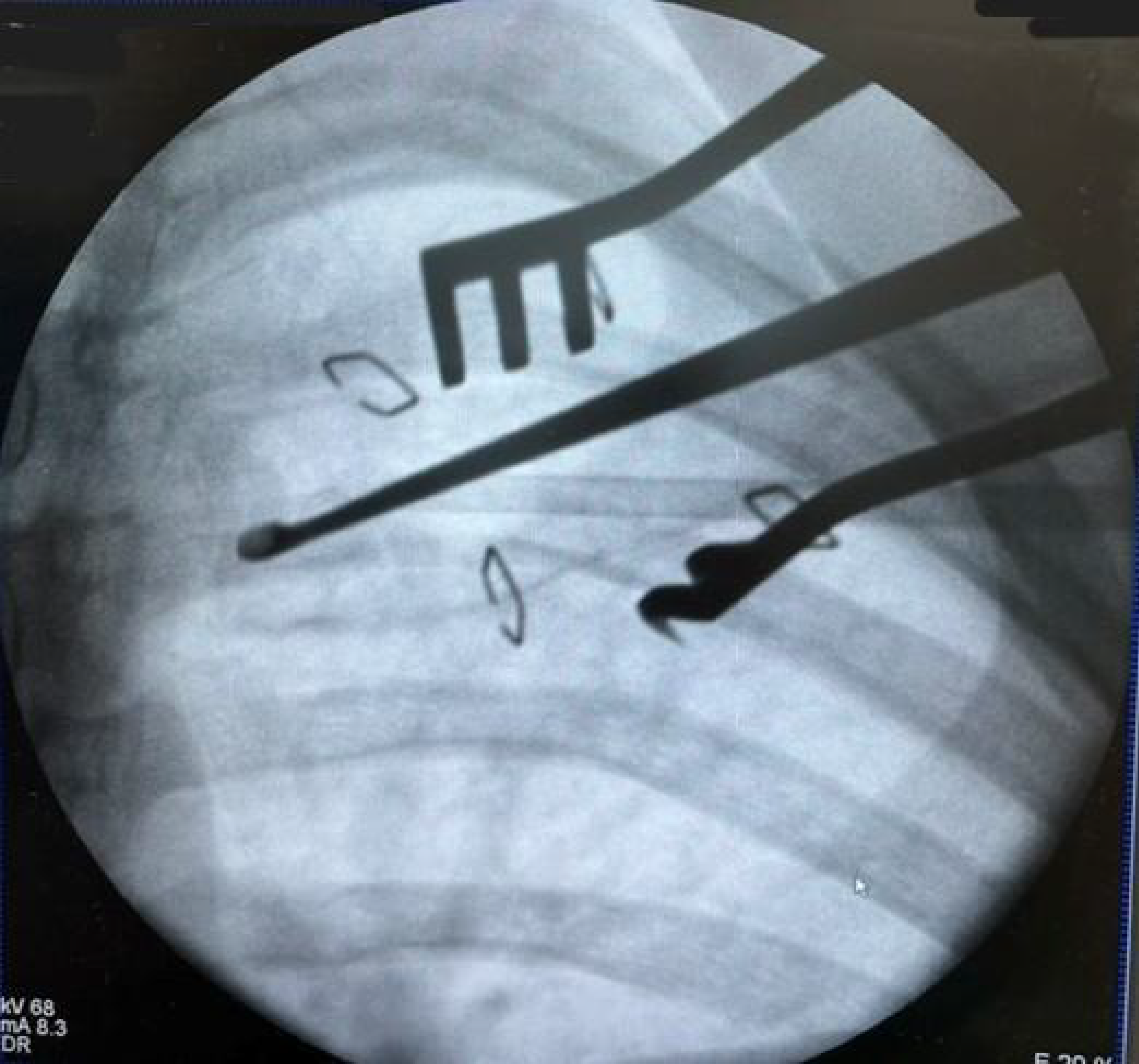
Figure 3
Image-guided open biopsy procedure.
- Citation: Gkougkoulias AN, Vlychou M, Dailiana ZH. Monostotic chronic nonbacterial osteomyelitis/chronic recurrent multifocal osteomyelitis of clavicle mimicking malignant bone tumor: A case report and review of literature. World J Clin Cases 2026; 14(22): 122012
- URL: https://www.wjgnet.com/2307-8960/full/v14/i22/122012.htm
- DOI: https://dx.doi.org/10.12998/wjcc.122012