
















Copyright: ©Author(s) 2026.
World J Clin Cases. Aug 6, 2026; 14(22): 121141
Published online Aug 6, 2026. doi: 10.12998/wjcc.121141
Published online Aug 6, 2026. doi: 10.12998/wjcc.121141
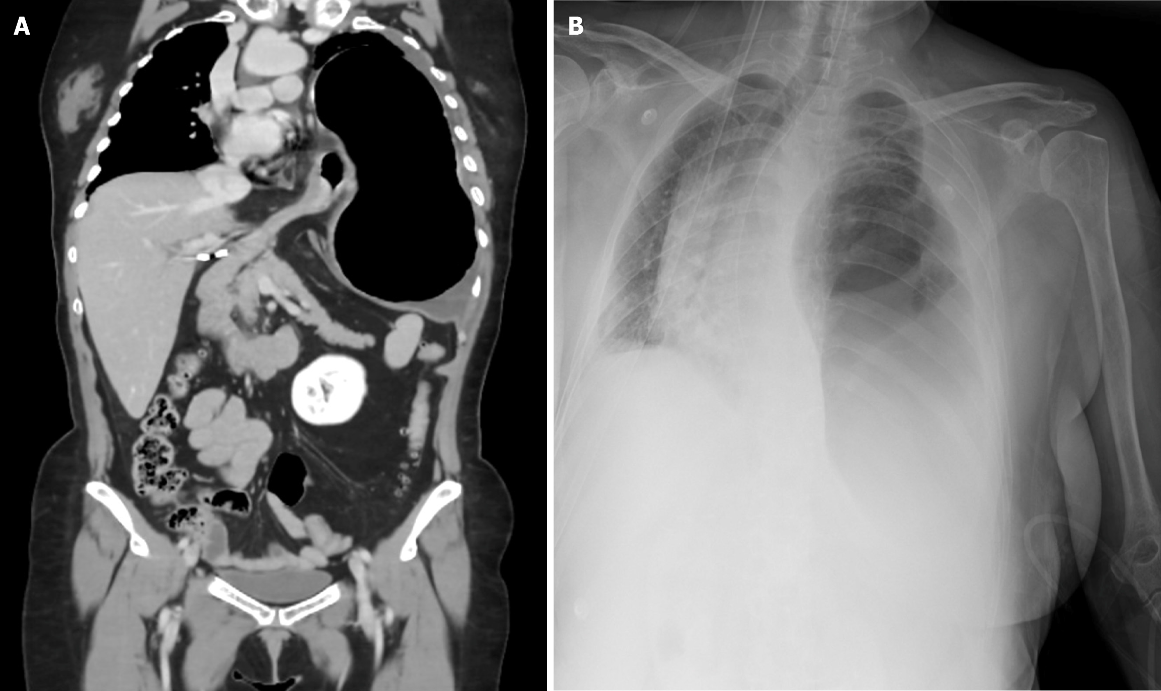
Figure 1 Initial emergency department imaging examinations (September 29th, 2025).
A: Contrast-enhanced computed tomography of the chest, abdomen, and pelvis demonstrating tension gastrothorax caused by a left paraesophageal hernia with intrathoracic migration of the stomach and proximal duodenum; B: Chest radiograph showing a complicated hiatal hernia with rightward displacement of mediastinal structures and a concomitant left pleural effusion.
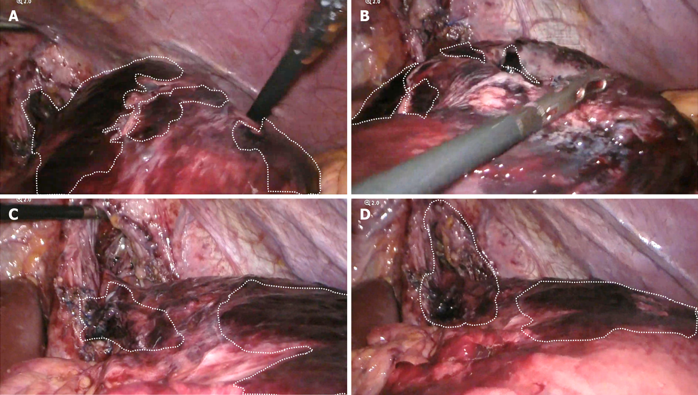
Figure 2 Intraoperative findings during initial laparoscopy.
A and B: Gastric body, fundus, and greater curvature demonstrating extensive ischemic changes and a large intramural hematoma; C: Ischemic involvement of the gastroesophageal junction; D: Ischemic changes affecting the gastric body.
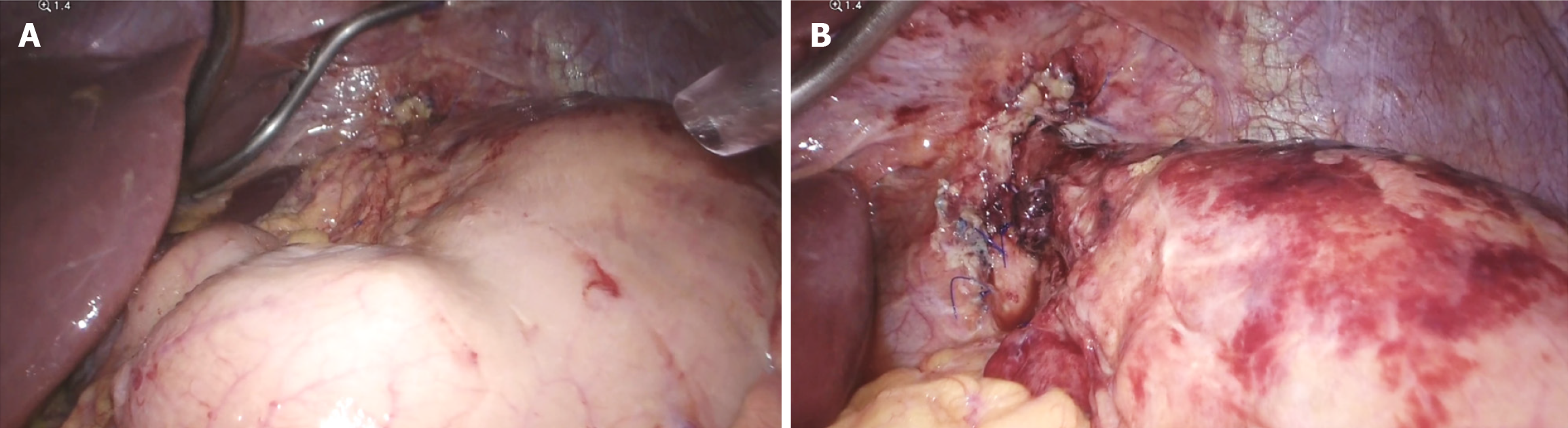
Figure 3 Intraoperative findings during second-look laparoscopy.
A: Gastric body demonstrating restoration of normal color and perfusion; B: Gastroesophageal junction and fundus showing resolving ischemic changes with minimal residual serosal erythema.
- Citation: Hagerman-Sucar G, Vázquez-Nicolás DA, Quiñones-Corona V, Olaya-Herrera M, Rodríguez Reyes A, Furuzawa-Carballeda J, Aguilar-León D, Torres-Villalobos G. Acute tension gastrothorax post hiatal hernia repair and Nissen fundoplication: A case report. World J Clin Cases 2026; 14(22): 121141
- URL: https://www.wjgnet.com/2307-8960/full/v14/i22/121141.htm
- DOI: https://dx.doi.org/10.12998/wjcc.121141