
















Copyright: ©Author(s) 2026.
World J Clin Cases. Aug 6, 2026; 14(22): 120306
Published online Aug 6, 2026. doi: 10.12998/wjcc.120306
Published online Aug 6, 2026. doi: 10.12998/wjcc.120306
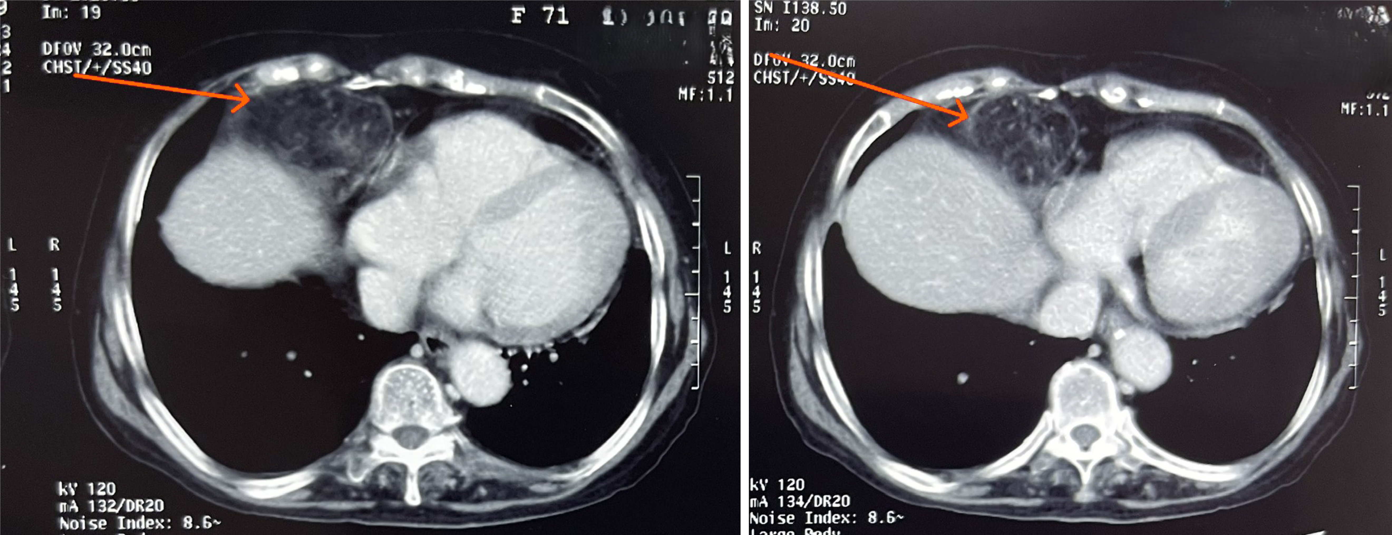
Figure 1
Computed tomography of the hernia through the right hemidiaphragm (arrows).
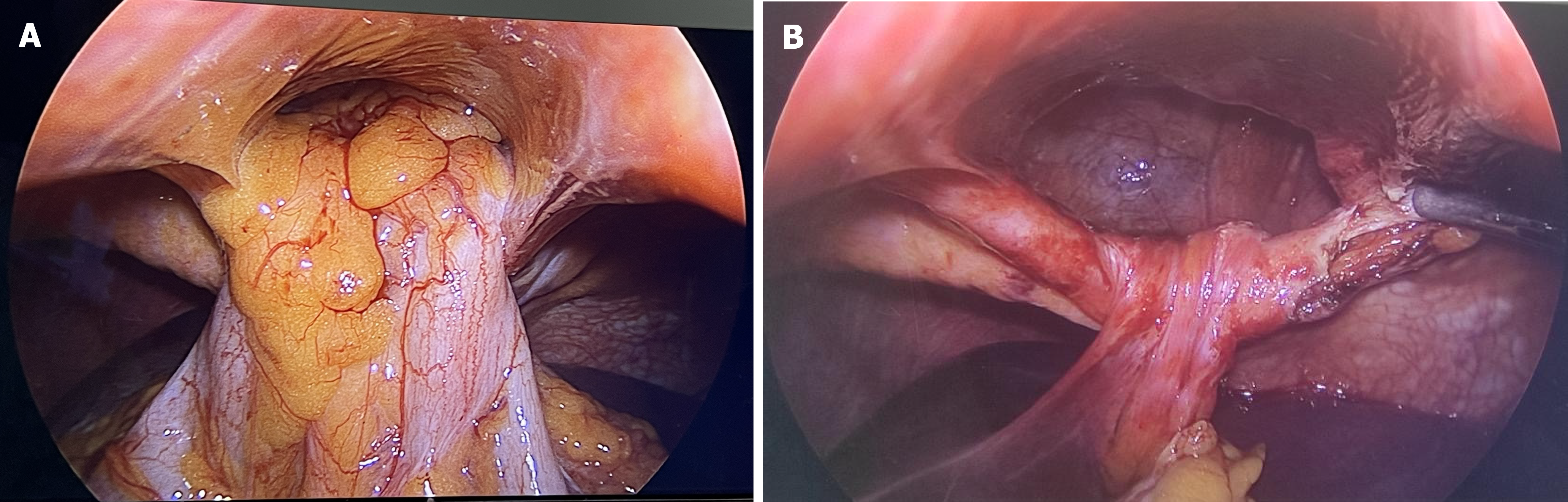
Figure 2 Intraoperative view of the hernia.
A: Transverse colon and omentum herniation through the Morgagni hernia defect; B: Morgagni hernia defect after the release of the colon and omentum in the abdominal cavity.
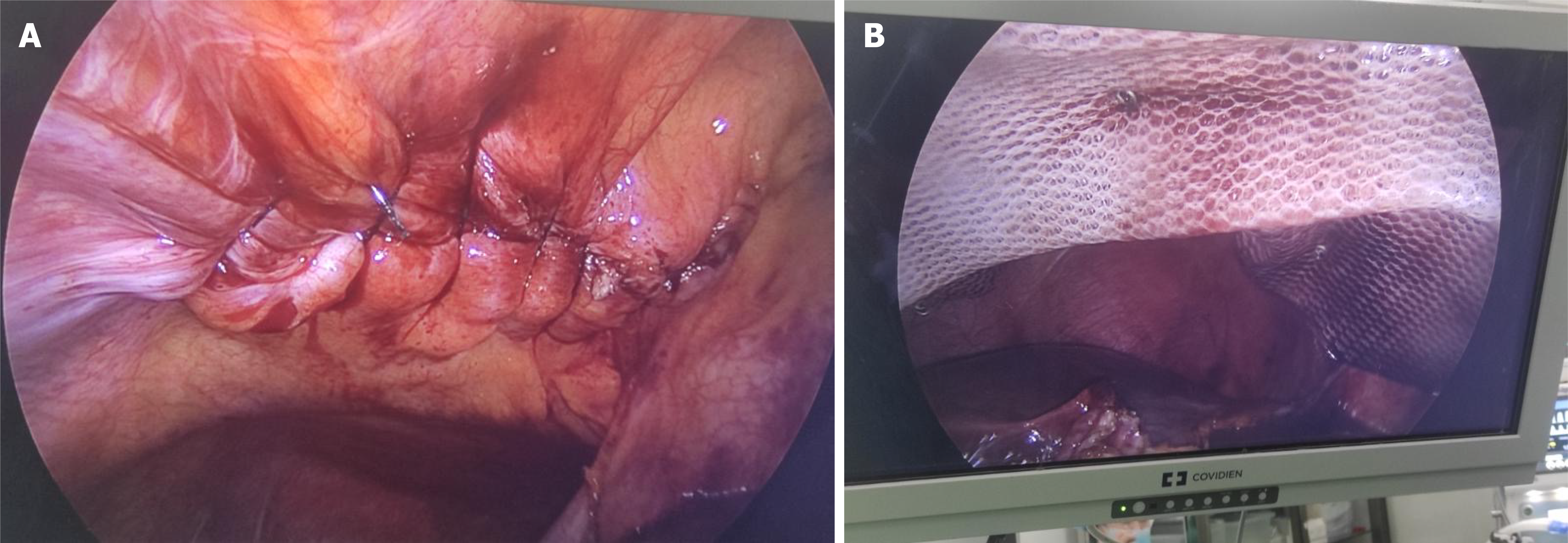
Figure 3 Treatment.
A: Repair of the defect using single Prolene 10 sutures; B: Reinforcement was achieved with a 15 cm × 20 cm Parietex™ mesh secured in place using Protack™ fixation devices.
- Citation: Nguyen HN. Laparoscopic repair of a rare Morgagni hernia: A case report. World J Clin Cases 2026; 14(22): 120306
- URL: https://www.wjgnet.com/2307-8960/full/v14/i22/120306.htm
- DOI: https://dx.doi.org/10.12998/wjcc.120306