
















Copyright: ©Author(s) 2026.
World J Clin Cases. Jul 26, 2026; 14(21): 120561
Published online Jul 26, 2026. doi: 10.12998/wjcc.120561
Published online Jul 26, 2026. doi: 10.12998/wjcc.120561
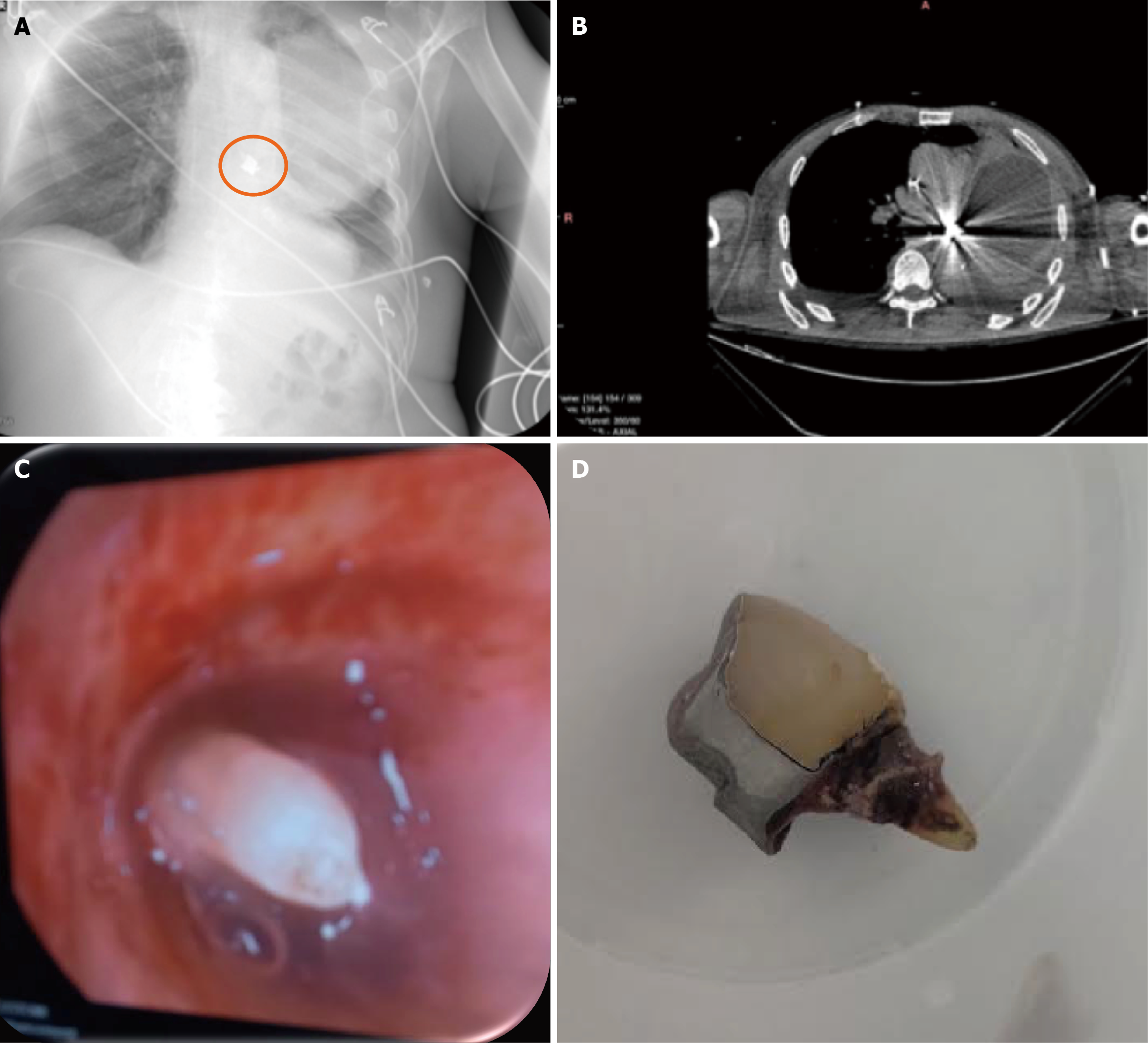
Figure 1 Imaging results.
A: Post-tracheostomy chest radiograph demonstrating a sharply marginated radiopaque foreign body projected over the region of the left main bronchus (within the orange circle); B: Chest computed tomography showing a dense endobronchial object within the proximal left main bronchus. The high radiodensity of the object with associated beam-hardening artifacts limited precise morphological characterization on imaging alone; C: Flexible bronchoscopic view revealing a natural tooth with an attached metallic crown partially embedded in the left main bronchus; D: Extracted foreign body consisting of a natural tooth with a metallic crown following successful removal by flexible bronchoscopy using tripod grasping forceps.
- Citation: Karadimou K, Mavrovounis G, Karapetsa M, Pantazopoulos I. Aspiration of a metallic-crowned tooth in the left main bronchus in a neurocritical patient: A case report. World J Clin Cases 2026; 14(21): 120561
- URL: https://www.wjgnet.com/2307-8960/full/v14/i21/120561.htm
- DOI: https://dx.doi.org/10.12998/wjcc.120561