
















Copyright: ©Author(s) 2026.
World J Clin Cases. Jul 16, 2026; 14(20): 122343
Published online Jul 16, 2026. doi: 10.12998/wjcc.122343
Published online Jul 16, 2026. doi: 10.12998/wjcc.122343
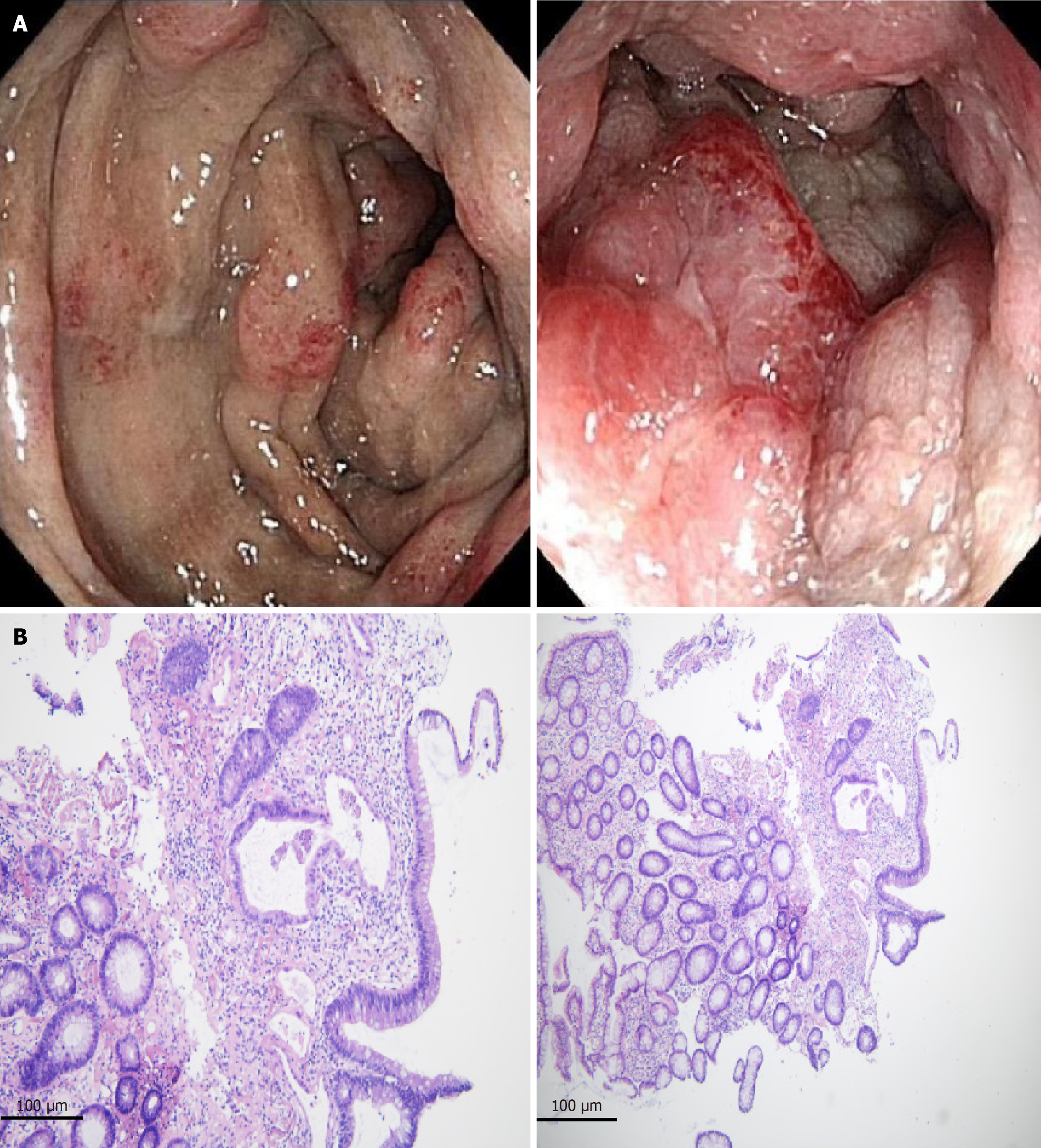
Figure 1 Endoscopic and histopathological findings.
A: Colonoscopy showing nodular swelling, bluish-purple mucosa, mucosal friability, and luminal narrowing; B: Histopathological examination showing tubular adenoma-like epithelial changes without definite evidence of invasive malignancy.
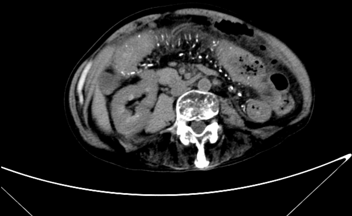
Figure 2 Abdominal computed tomography findings.
Abdominal computed tomography showing diffuse colonic wall thickening and extensive linear calcifications along the mesenteric veins and colonic wall, findings characteristic of idiopathic mesenteric phlebosclerosis.
- Citation: Li BJ, Duan CY, Wang YS, Ma XP, Sun LX. Idiopathic mesenteric phlebosclerosis associated with long-term herbal medicine use: A case report. World J Clin Cases 2026; 14(20): 122343
- URL: https://www.wjgnet.com/2307-8960/full/v14/i20/122343.htm
- DOI: https://dx.doi.org/10.12998/wjcc.122343