
















Copyright: ©Author(s) 2026.
World J Clin Cases. Jul 16, 2026; 14(20): 121215
Published online Jul 16, 2026. doi: 10.12998/wjcc.121215
Published online Jul 16, 2026. doi: 10.12998/wjcc.121215
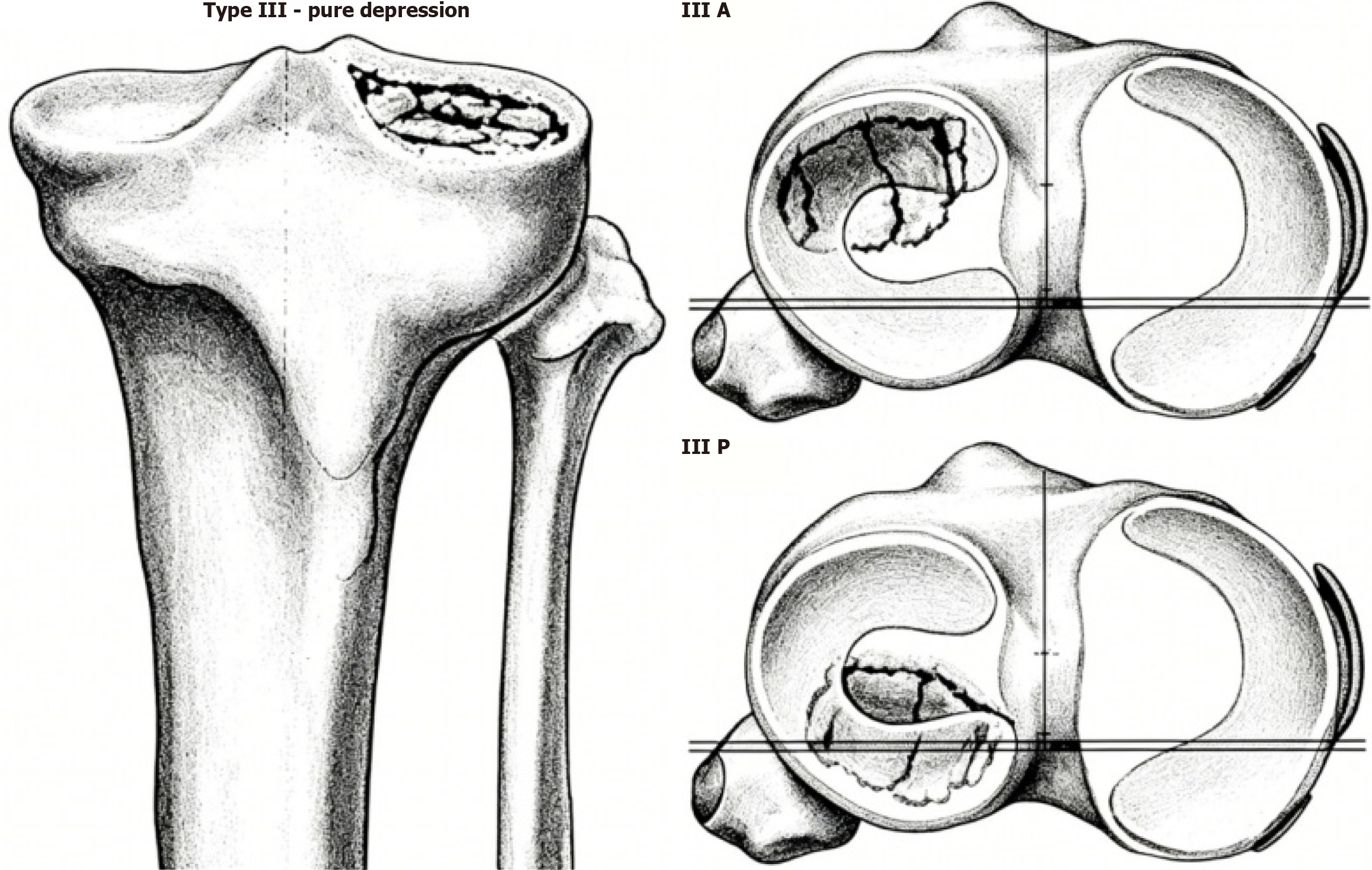
Figure 1 Schatzker type 3 fractures.
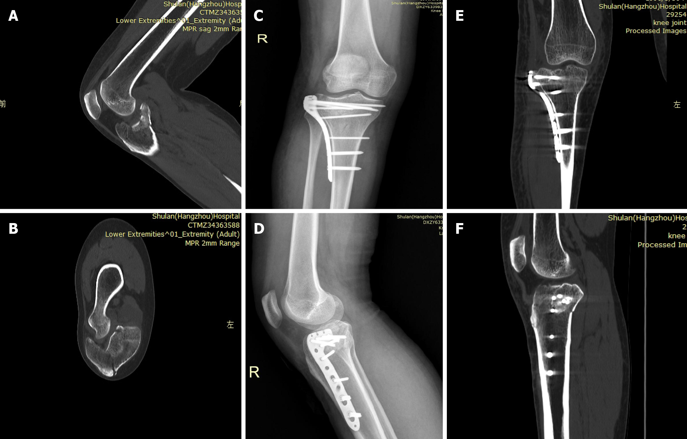
Figure 2 Postoperative follow-up imaging.
A: Preoperative computed tomography (CT) sagittal scan of tibial plateau showing a posterior tibial plateau depression fracture; B: Preoperative coronal CT scan of the tibial plateau showing a depressed fracture on the lateral side of the tibial plateau; C: Postoperative follow-up X-ray positive position film. D: Postoperative follow-up X-ray lateral view film; E: Postoperative follow-up CT coronal scan; F: Postoperative follow-up CT sagittal images (C-F: Postoperative follow-up X-ray and CT scan showing healing of the fractures and osteotomy blocks). There was no bone collapse, nonunion, or infection.
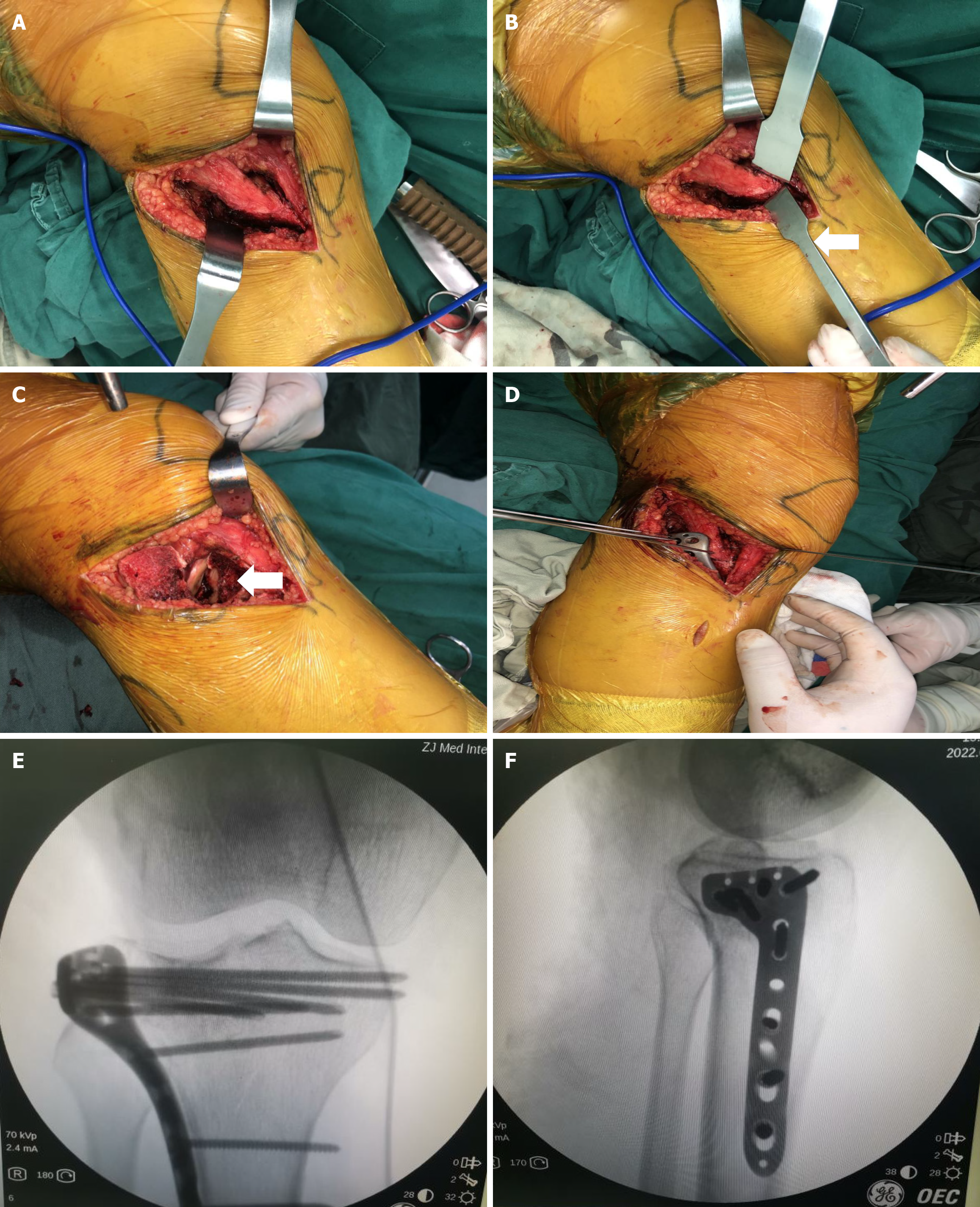
Figure 3 Surgical procedure and intraoperative imaging.
A: Exposure of the iliotibial tract; B: Use of a thin osteotome to make an inverted L-shaped osteotomy in front of and below the Gerdy tubercle (arrow); C: Lifting the osteotomy block together with the iliotibial tract upward and backward, clearly showing the depressed fracture (arrow); D: Fixation with Kirschner wires, and placement of an L-shaped locking anatomical plate on the anterior-lateral side of the proximal tibia; E and F: Under fluoroscopy, the fracture reduction is satisfactory and the internal fixation is appropriate.
- Citation: Lin DC, Hu TY, Zhou YJ, Zhang ZW, Yuan JJ, Lu CX, Zheng LN. Modified Gerdy tubercle osteotomy in the treatment of posterolateral tibial plateau depression fractures: Technical report and preliminary results. World J Clin Cases 2026; 14(20): 121215
- URL: https://www.wjgnet.com/2307-8960/full/v14/i20/121215.htm
- DOI: https://dx.doi.org/10.12998/wjcc.121215