Copyright
©The Author(s) 2025.
World J Clin Cases. Mar 26, 2025; 13(9): 99463
Published online Mar 26, 2025. doi: 10.12998/wjcc.v13.i9.99463
Published online Mar 26, 2025. doi: 10.12998/wjcc.v13.i9.99463
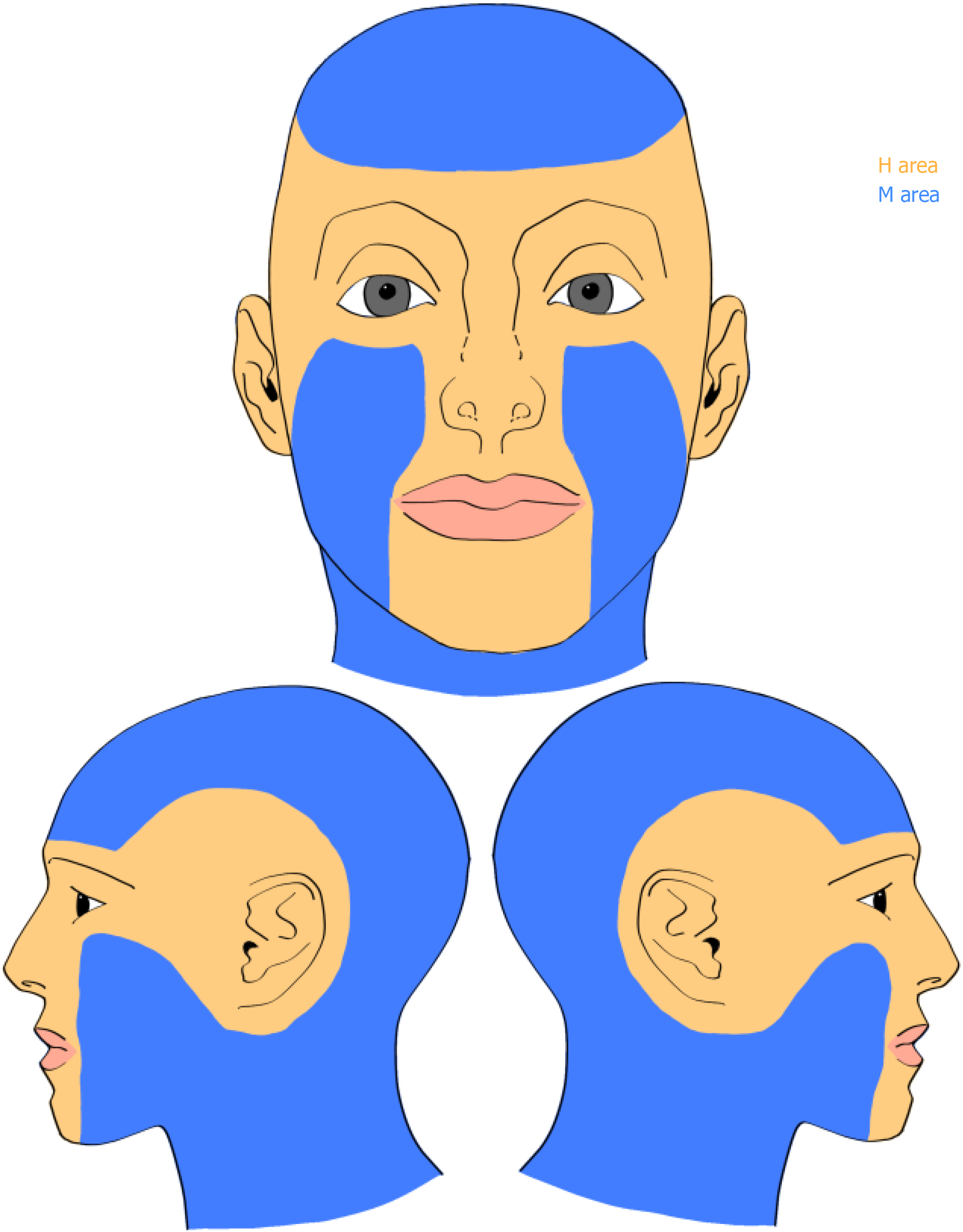
Figure 1 Classification of tumor location into high-risk and mid-risk zones.
Area H (yellow area) encompasses central facial regions, including eyelids, eyebrows, periorbital area, nose, lips, chin, mandible, preauricular, and postauricular areas; area M (blue area) includes the cheeks, forehead, and scalp.
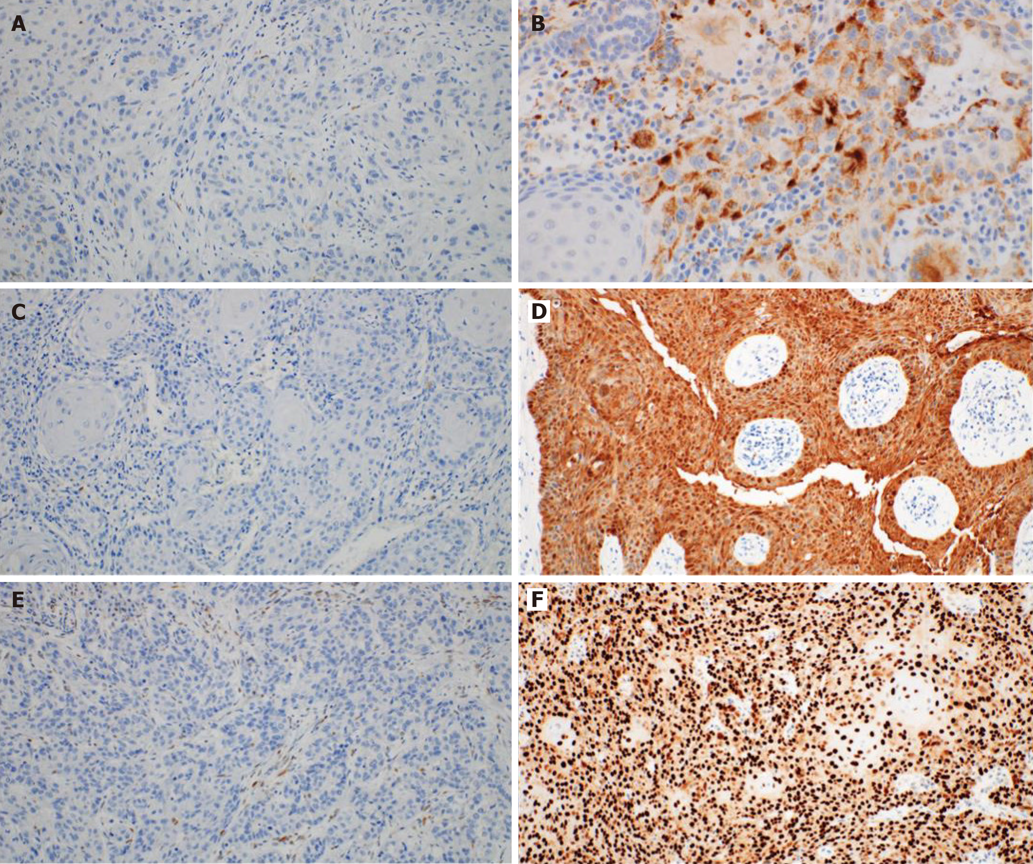
Figure 2 Representative images of immunohistochemical study are shown.
A: Images of human papillomavirus staining, negative (× 200); B: Images of human papillomavirus staining, positive (× 400); C: Images of p16 staining, negative (× 200); D: Images of p16 staining, positive (× 200); E: Images of p53 staining, negative (× 200); F: Images of p53 staining, positive (× 200).
- Citation: Nam HJ, Ryu H, Lee DW, Byeon JY, Kim JH, Lee JH, Lim S, Choi HJ. Expression rates of p16, p53 in head and neck cutaneous squamous cell carcinoma based on human-papillomavirus positivity. World J Clin Cases 2025; 13(9): 99463
- URL: https://www.wjgnet.com/2307-8960/full/v13/i9/99463.htm
- DOI: https://dx.doi.org/10.12998/wjcc.v13.i9.99463


