©The Author(s) 2025.
World J Clin Cases. Nov 6, 2025; 13(31): 108942
Published online Nov 6, 2025. doi: 10.12998/wjcc.v13.i31.108942
Published online Nov 6, 2025. doi: 10.12998/wjcc.v13.i31.108942
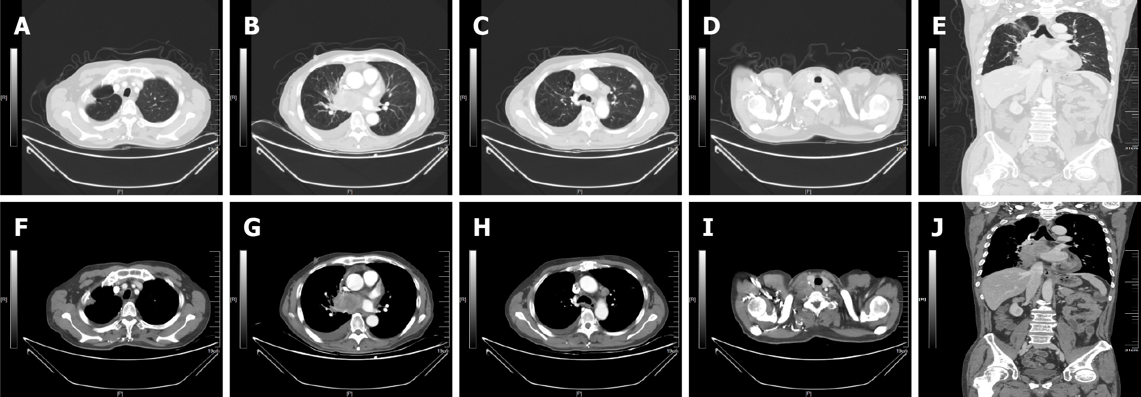
Figure 1 Contrast-enhanced computed tomography images.
A-E: Lung window; A: The images show the local nodular thickening of the right pleura; B: The mass film shadow near the right lung portal; C: The images show the pleural effusion; D: The images show the cervical lymph nodes metastasis; E: Coronal observation shows that the mediastinal lymph nodes are enlarged; F-J: Mediastinal window; F: The images show the local nodular thickening of the right pleura; G: The mass film shadow near the right lung portal; H: The images show the pleural effusion; I: The images show the cervical lymph nodes metastasis; J: Coronal observation shows that the mediastinal lymph nodes are enlarged.
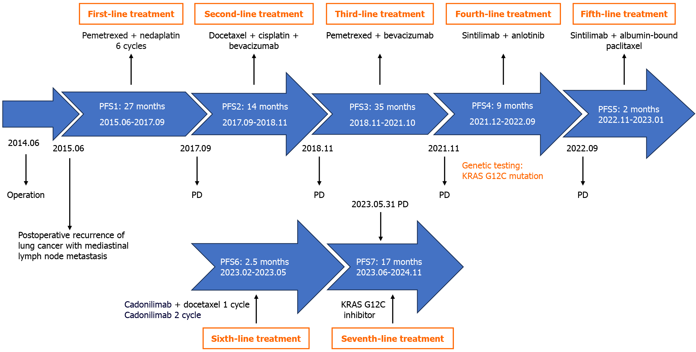
Figure 2 Treatment course of the patient with lung adenocarcinoma.
PD: Progressive disease; PFS: Progression-free survival; KRAS G12C: Kirsten rat sarcoma viral oncogene homolog at glycine 12 to cysteine.
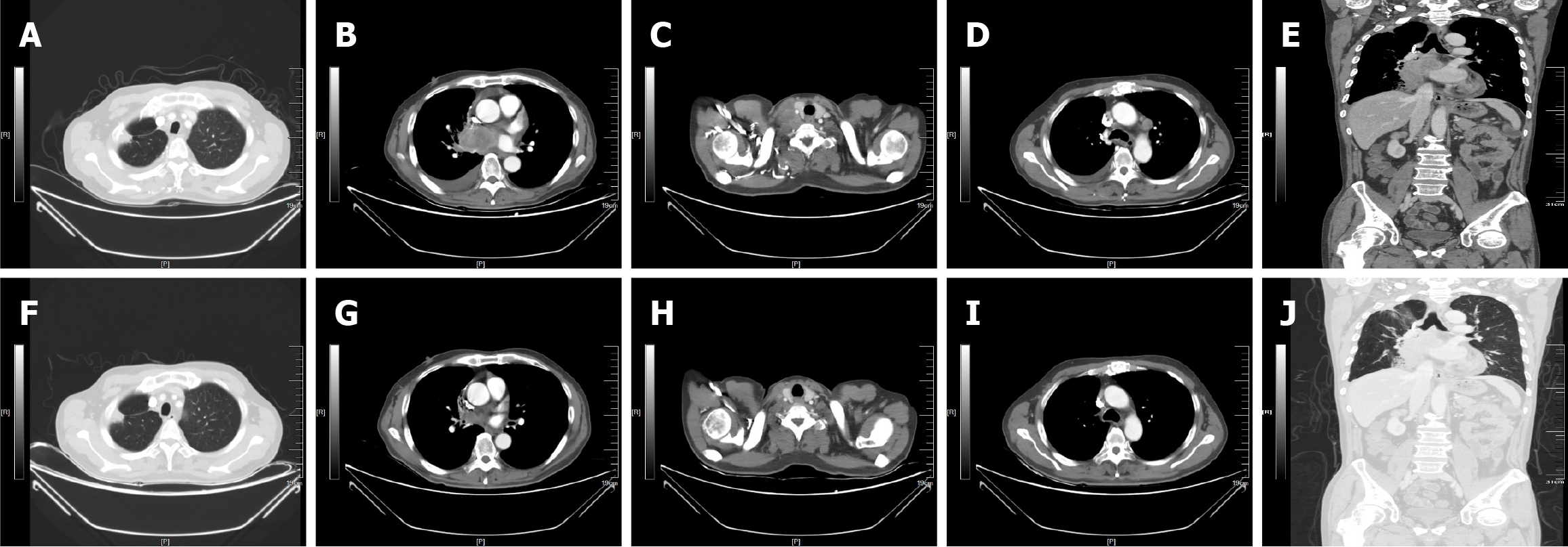
Figure 3 Representative radiologic images of the patient before and after the Kirsten rat sarcoma viral oncogene homolog at glycine 12 to cysteine inhibitor.
A-E: May 31, 2023; F-J: July 25, 2023.
Figure 4 Enhanced computed tomography showed the follow-up of the left diaphragmatic angle lymph node.
A: On May 31, 2023; B: On May 7, 2024; C: On July 10, 2024; D: On September 11, 2024; E: On November 11, 2024. The orange arrows in the figures point to the metastatic left diaphragmatic angle lymph node.
- Citation: Gan L, Shen JF, Yao MX, Chen ZG, Zhuang ZX. Kirsten rat sarcoma G12C inhibitor treatment for a patient with relapsed metastatic lung adenocarcinoma: A case report. World J Clin Cases 2025; 13(31): 108942
- URL: https://www.wjgnet.com/2307-8960/full/v13/i31/108942.htm
- DOI: https://dx.doi.org/10.12998/wjcc.v13.i31.108942


