©The Author(s) 2025.
World J Clin Cases. Nov 6, 2025; 13(31): 108301
Published online Nov 6, 2025. doi: 10.12998/wjcc.v13.i31.108301
Published online Nov 6, 2025. doi: 10.12998/wjcc.v13.i31.108301
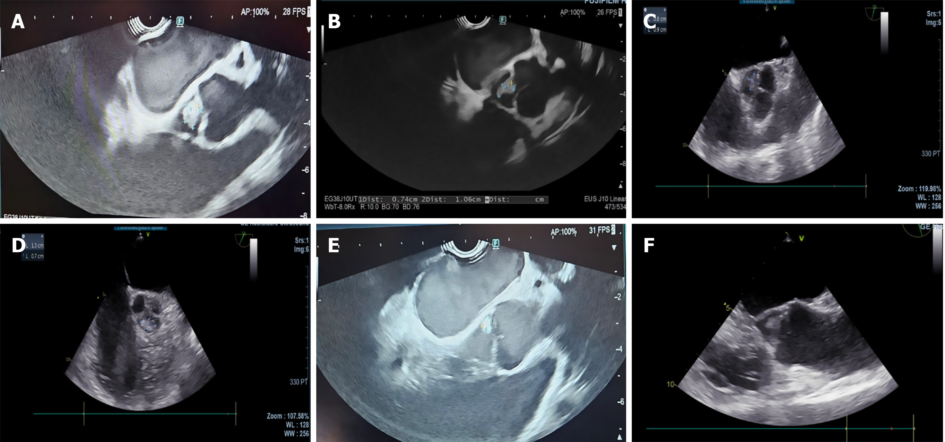
Figure 1 Computed tomography view of the aortic valve lesion.
A: Endoscopic ultrasound longitudinal; B: Endoscopic ultrasound longitudinal; C: Transesophageal echocardiography cross-sectional; D: Transesophageal echocardiography cross-sectional; E: Transesophageal echocardiography longitudinal; F: Endoscopic ultrasound longitudinal.
- Citation: Elsayed G, Mohamed L, Almasaabi M, Barakat K, Taha R, AlQahtani MS, Makdisi G, Musa M, Alfadda A, Gadour E. Incidental detection of aortic valve fibroelastomas during endoscopic ultrasound for pancreatic evaluation: Three case reports. World J Clin Cases 2025; 13(31): 108301
- URL: https://www.wjgnet.com/2307-8960/full/v13/i31/108301.htm
- DOI: https://dx.doi.org/10.12998/wjcc.v13.i31.108301


