©The Author(s) 2025.
World J Clin Cases. Oct 6, 2025; 13(28): 109536
Published online Oct 6, 2025. doi: 10.12998/wjcc.v13.i28.109536
Published online Oct 6, 2025. doi: 10.12998/wjcc.v13.i28.109536
Figure 1 Sagittal T2-weighted magnetic resonance imaging revealing an enlarged uterus with heterogeneous myometrial signal intensity and multiple cystic spaces.
A large, irregular uterine mass containing hemorrhagic components is observed, along with disruptions in myometrial continuity suggestive of possible rupture. The lesion exhibits areas of high signal intensity consistent with hemorrhage and necrosis. These findings raise concerns for an aggressive uterine malignancy.
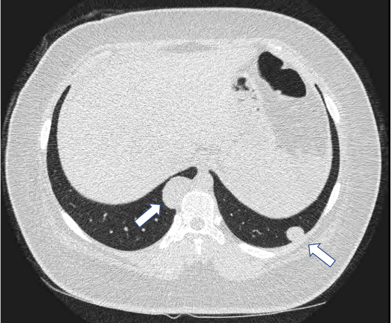
Figure 2 Axial chest computed tomography scan shows two well-defined, round pulmonary nodules (indicated by arrows) located in the lower lobes of both lungs.
These nodules were initially suspicious for metastatic lesions but were later confirmed to be endometriotic implants.
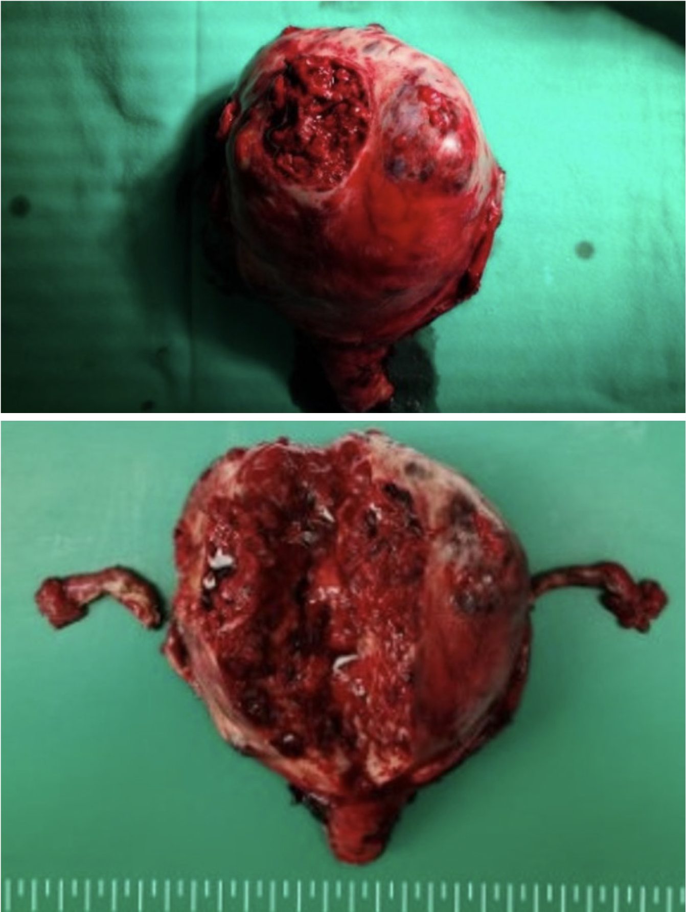
Figure 3 Gross pathology of a ruptured uterus due to severe adenomyosis with hemorrhagic necrosis following total hysterectomy and bilateral salpingo-oophorectomy.
The external surface of the uterus displays a large, irregularly necrotic area with extensive hemorrhage and tissue disruption. A bisected view of the excised uterus reveals diffuse hemorrhagic necrosis extending throughout the myometrium. The fallopian tubes appear intact, and no gross abnormalities are observed in the adnexa.
- Citation: Ju UC, Kang WD, Kim SM. Adenomyosis-associated uterine rupture and pulmonary endometriosis mimicking advanced-stage uterine malignancy in an adolescent female: A case report. World J Clin Cases 2025; 13(28): 109536
- URL: https://www.wjgnet.com/2307-8960/full/v13/i28/109536.htm
- DOI: https://dx.doi.org/10.12998/wjcc.v13.i28.109536


