©The Author(s) 2025.
World J Clin Cases. Oct 6, 2025; 13(28): 106852
Published online Oct 6, 2025. doi: 10.12998/wjcc.v13.i28.106852
Published online Oct 6, 2025. doi: 10.12998/wjcc.v13.i28.106852
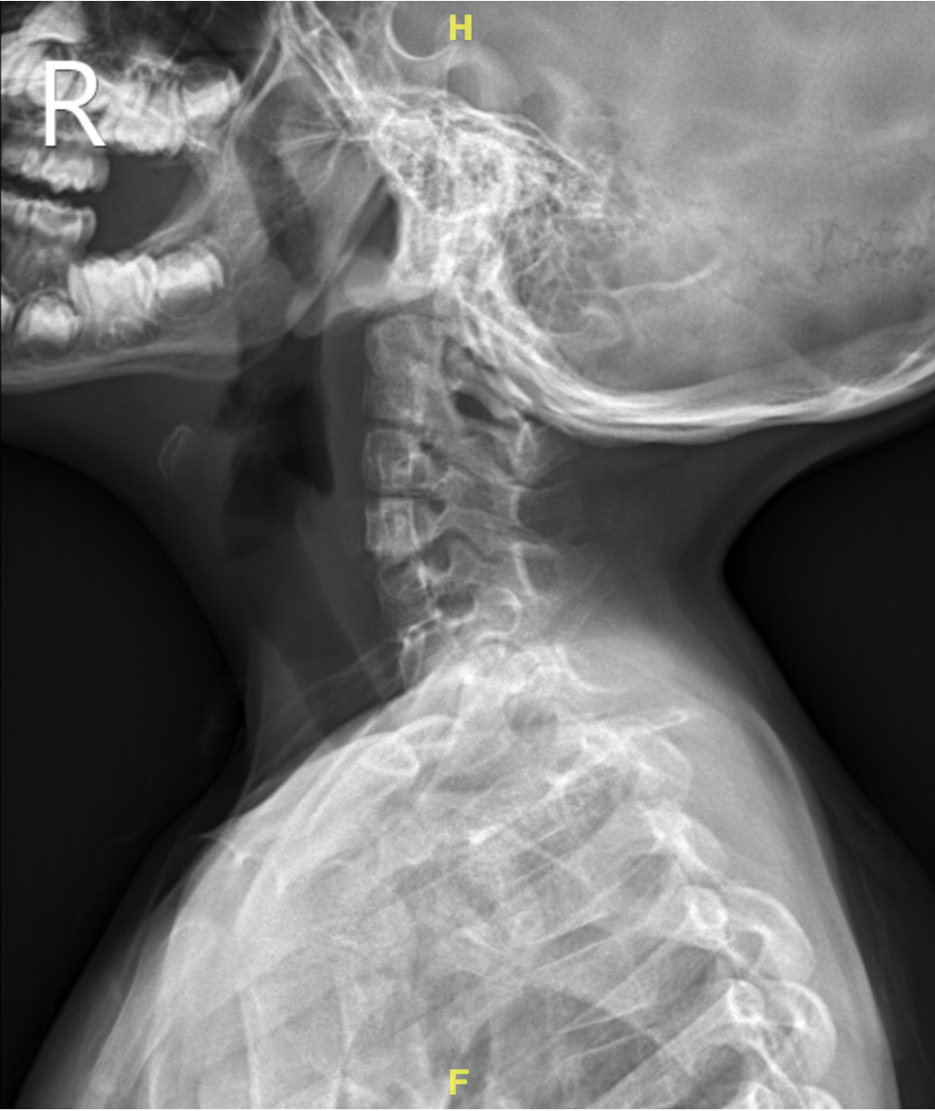
Figure 1 Pre-operative cervical spine X-ray.
The cervical spine X-ray showed that the alignment was within normal limits.
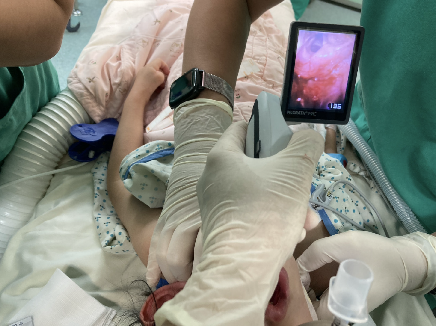
Figure 2 Laryngeal view under video laryngoscope.
The image from the McGRATH showed that the laryngeal view was classified as Cormack-Lehane grade 2
- Citation: Lin XN, Chan WS, Lu CW. Airway management strategies in a pediatric patient with MURCS association: A case report. World J Clin Cases 2025; 13(28): 106852
- URL: https://www.wjgnet.com/2307-8960/full/v13/i28/106852.htm
- DOI: https://dx.doi.org/10.12998/wjcc.v13.i28.106852


