©The Author(s) 2025.
World J Clin Cases. Sep 26, 2025; 13(27): 108693
Published online Sep 26, 2025. doi: 10.12998/wjcc.v13.i27.108693
Published online Sep 26, 2025. doi: 10.12998/wjcc.v13.i27.108693
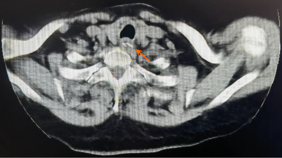
Figure 1 Computed tomography of the neck.
Foreign bodies can be seen in the upper digestive tract (orange arrow).
Figure 2 Esophageal endoscopy at different sites in the patient.
A: Esophageal endoscopy of the throat; B: Esophageal endoscopy 14 cm from the incisors, showing esophageal mucosal erosion; C: Esophageal endoscopy of the inlet patch; D: Esophageal endoscopy of food debris and a foreign body bone lodged in the esophagus.
- Citation: Qiao HW, Ye YF, Nie LX, Bai S, Du GZ. Disappearing intraesophageal foreign body: A case report. World J Clin Cases 2025; 13(27): 108693
- URL: https://www.wjgnet.com/2307-8960/full/v13/i27/108693.htm
- DOI: https://dx.doi.org/10.12998/wjcc.v13.i27.108693


