©The Author(s) 2025.
World J Clin Cases. Sep 26, 2025; 13(27): 108261
Published online Sep 26, 2025. doi: 10.12998/wjcc.v13.i27.108261
Published online Sep 26, 2025. doi: 10.12998/wjcc.v13.i27.108261
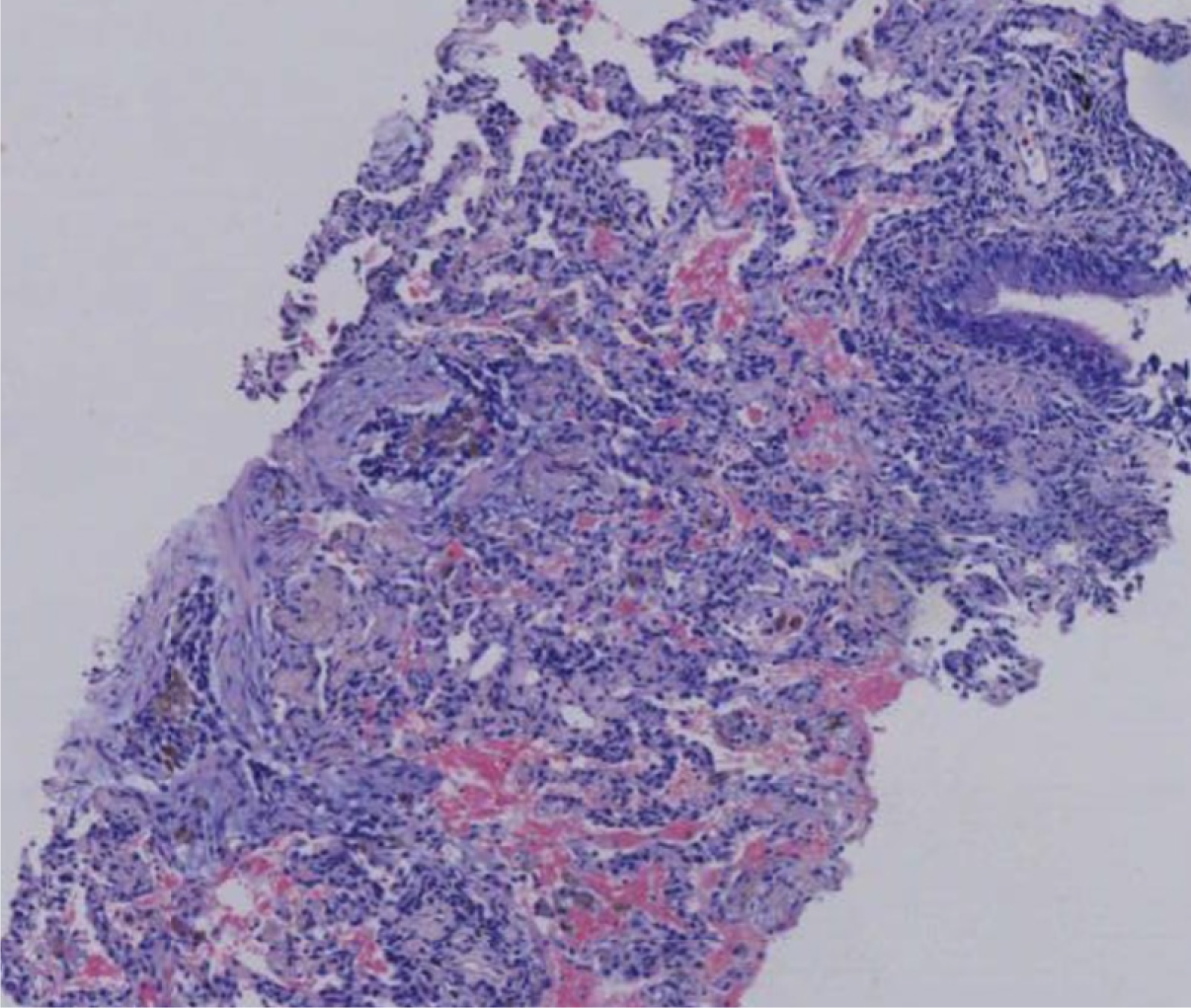
Figure 1 Pathological assessment showed localized fibrous tissue hyperplasia and extensive plasma cell infiltration.
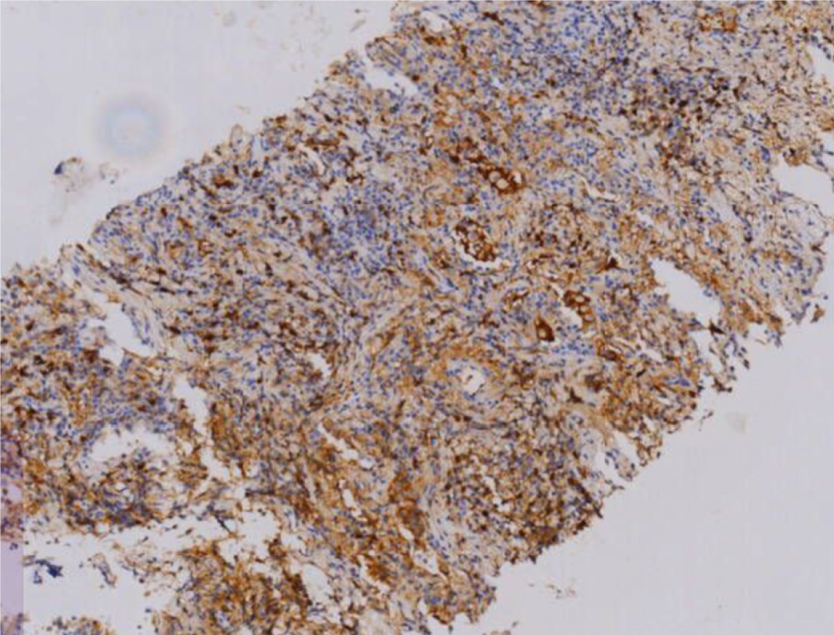
Figure 2 A total of 30-40 immunoglobulin G4-positive cells per high-power field, and an immunoglobulin G4/immunoglobulin G ratio of 40%.
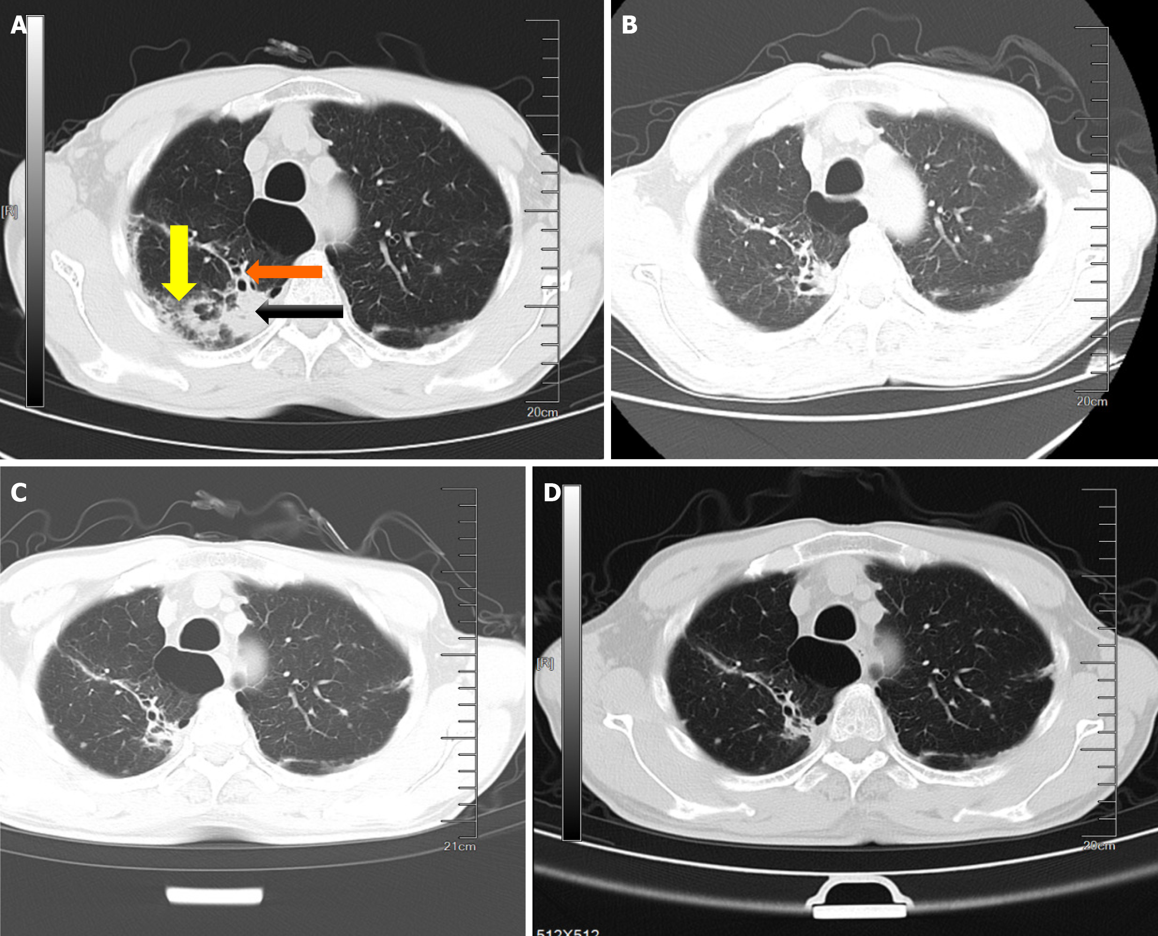
Figure 3 Axial unenhanced thoracic computed tomography.
A: Before treatment. Showing distal bronchial distension and traction bronchiectasis (orange arrow), glass opacities (yellow arrow) and a mass in the right superior lobe (black arrow); B: Computed tomography (CT) scan showing that the mass had shrunk and the ground-glass lesions had cleared after 2 months of corticosteroid therapy (35 mg/day oral prednisolone and mycophenolate mofetil 0.5 g bid); C: Chest CT scan at 6 months of treatment showing that the mass had shrunk further; D: Chest CT obtained 1 year after treatment. Chest CT shows that only cord-like shadows remained.
- Citation: Zhou JL, Zhou XY, Li WJ, Feng S. Immunoglobulin G4-related lung disease mistaken for pulmonary tuberculosis: A case report. World J Clin Cases 2025; 13(27): 108261
- URL: https://www.wjgnet.com/2307-8960/full/v13/i27/108261.htm
- DOI: https://dx.doi.org/10.12998/wjcc.v13.i27.108261


