©The Author(s) 2025.
World J Clin Cases. Sep 16, 2025; 13(26): 104421
Published online Sep 16, 2025. doi: 10.12998/wjcc.v13.i26.104421
Published online Sep 16, 2025. doi: 10.12998/wjcc.v13.i26.104421
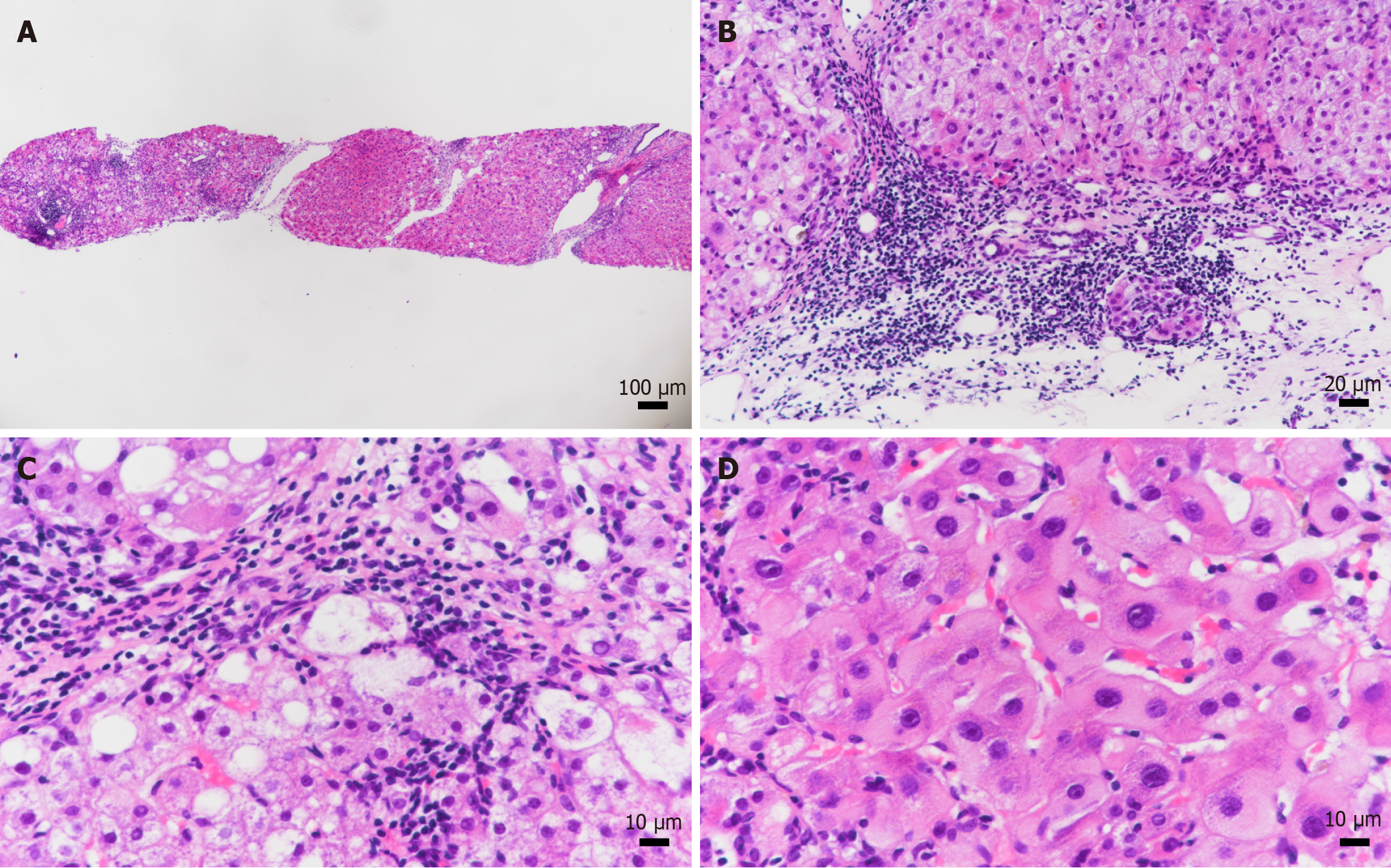
Figure 1 Liver biopsy pathological results for case 1.
A: A total of eleven small and medium-sized confluent areas were seen in the perforated liver tissue, with disorganized lobular architecture (hematoxylin and eosin); B: The main lesions were enlargement of most of the confluent areas, moderate inflammatory cell infiltration, predominantly single nucleated cells, and plasma cells were seen, often forming foci of lymphocyte aggregation, and mild-moderate interfacial inflammation (hematoxylin and eosin); C: The small bile ducts were discernible, and the biliary epithelium was irregularly arranged, and a mild-moderate fine biliary reaction was seen in the periphery of the confluent areas; fibroblastic tissue hyperplasia was seen in the confluent area, and the fibrous intervals of the bridge were formed, which destroyed the hepatic lobules by separating the hepatic parenchyma. Hepatocellular lipoatrophy and ballooning were seen in the lobules (hematoxylin and eosin); D: With focally necrotic and apoptotic vesicles, and mild inflammatory cell infiltration in the sinusoids, with some hepatocytes with ground-glass lesions (hematoxylin and eosin).
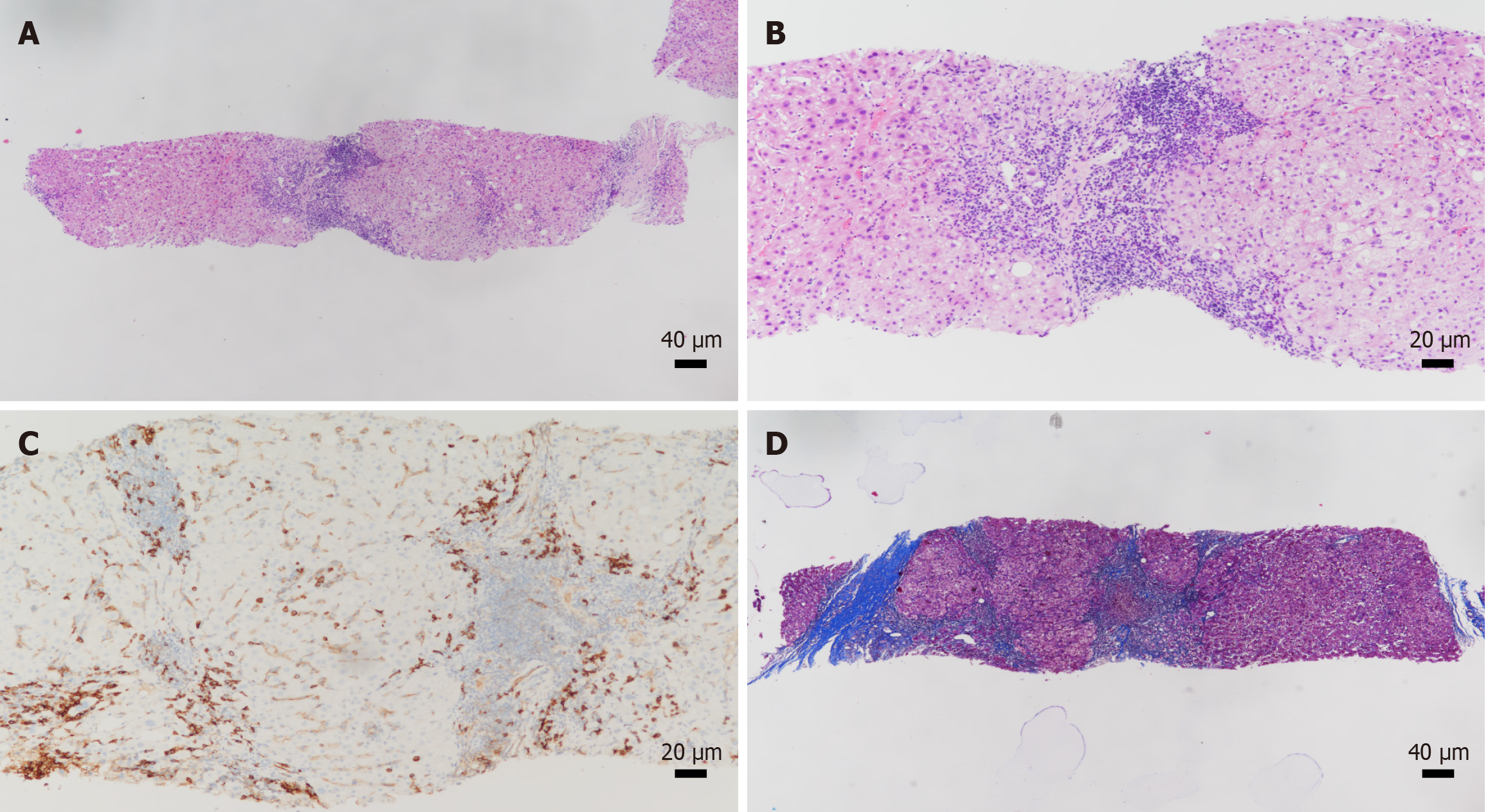
Figure 2 Liver biopsy pathological results for case 2.
A: The liver biopsy tissue revealed seven portal areas of medium to small size with disorganized lobular architecture (hematoxylin and eosin); B: The primary lesions include expansion of some portal areas, moderate inflammatory cell infiltration dominated by mononuclear cells (hematoxylin and eosin); C: Plasma cells are easily visible in small clusters (CD38), and moderate interface hepatitis observed in some portal areas; D: The bile ductules are discernible with disorganized epithelial lining and infiltration of inflammatory cells into the interductal epithelium, accompanied by a mild bile duct reaction around the portal areas. There is also proliferation of interstitial fibrous tissue in the portal areas, leading to the formation of local fibrous septa that segment the hepatic parenchyma and disrupt the lobular structure (Masson).
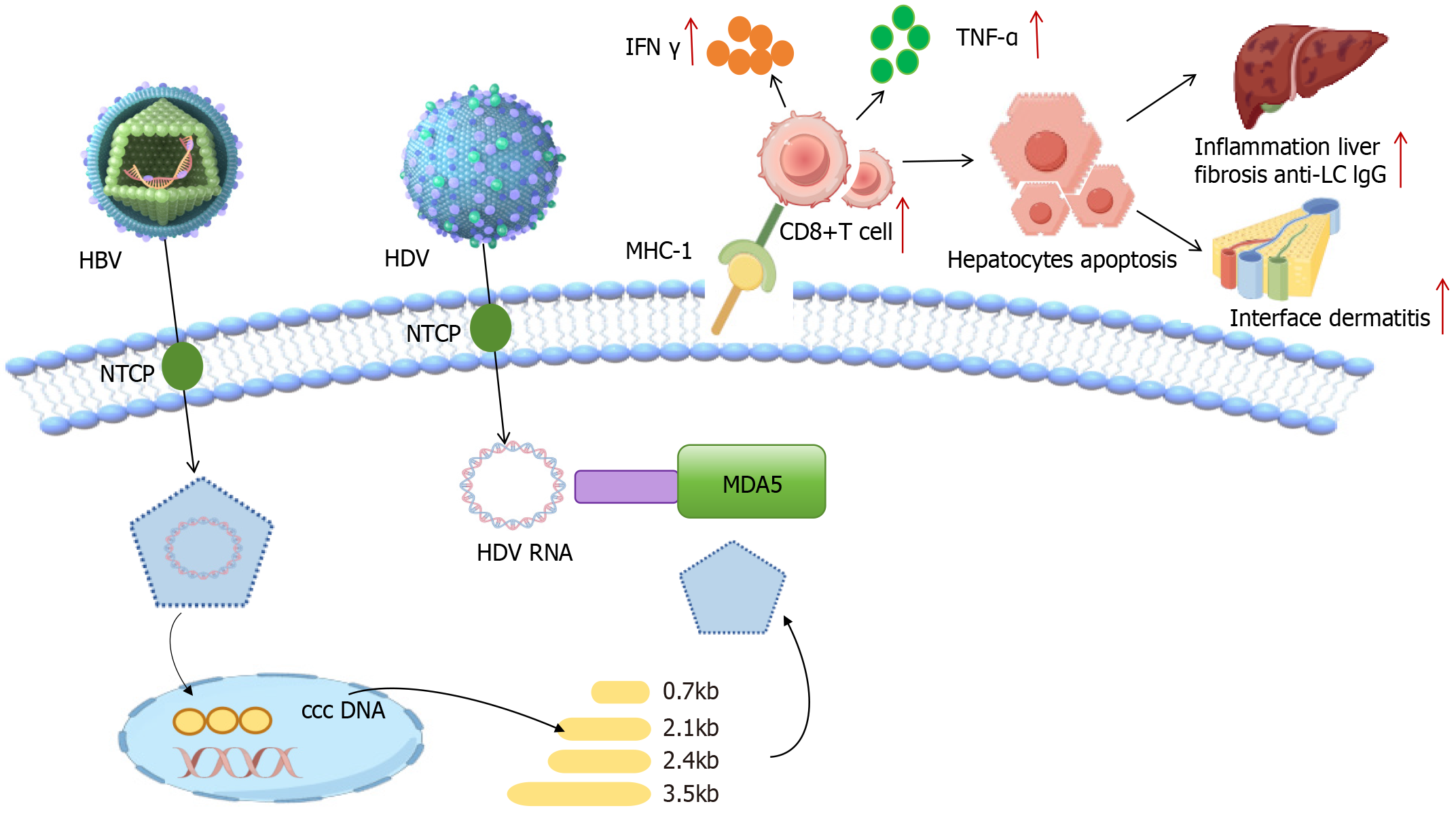
Figure 3 Hepatitis D virus, hepatitis B virus and autoimmune hepatitis interaction mechanism diagram.
HBV: Hepatitis B virus; HDV: Hepatitis D virus; IFN: Interferon; MHC: Major histocompatibility complex; TNF: Tumor necrosis factor; LC: Liver cancer; IgG: Immunoglobulin G; NTCP: Sodium taurocholate co-transporting polypeptide; MDA5: Melanoma differentiation-associated protein 5; cccDNA: Covalently closed circular DNA.
- Citation: Dou J, Zhao XY, Wang ZG, Ning ZH, Wang XZ, Guo F. Hepatitis B virus and hepatitis D virus co-infection complicated by autoimmune hepatitis: Two case reports. World J Clin Cases 2025; 13(26): 104421
- URL: https://www.wjgnet.com/2307-8960/full/v13/i26/104421.htm
- DOI: https://dx.doi.org/10.12998/wjcc.v13.i26.104421


