©The Author(s) 2025.
World J Clin Cases. Sep 6, 2025; 13(25): 107471
Published online Sep 6, 2025. doi: 10.12998/wjcc.v13.i25.107471
Published online Sep 6, 2025. doi: 10.12998/wjcc.v13.i25.107471
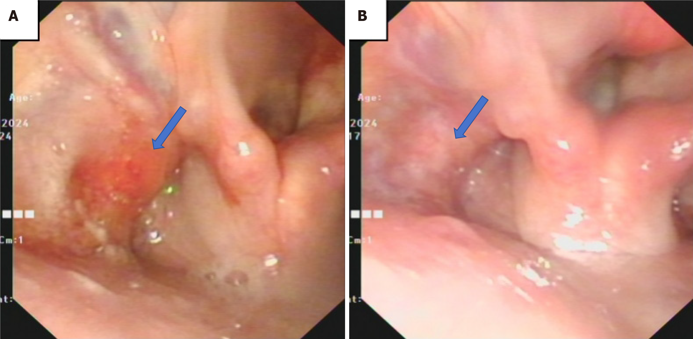
Figure 1 The laryngoscopy in the patient.
A: Pre-treatment laryngoscopy: Blood-stained secretions attached to the lateral wall of the left piriform sinus, narrowing of the sinus; B: Laryngoscopy one month post-treatment: With disappearance of blood-stained secretions in the left piriform sinus, narrowing was less pronounced.
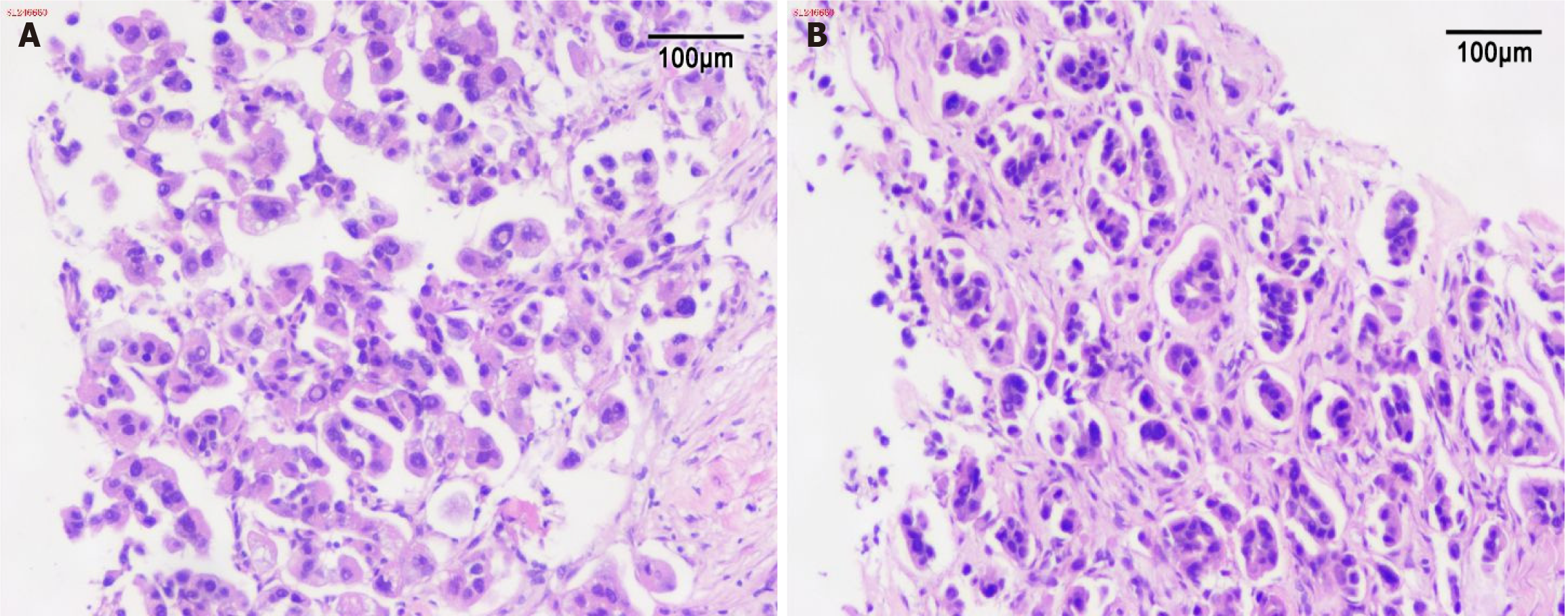
Figure 2 The pathology of oropharyngeal and abdominal wall biopsy.
The immunohistochemical staining results indicate: TTF-1 (+), Napsin A (+). A: (Laryngeal biopsy tissue) Microscopic examination reveals a small amount of striated muscle tissue and fibrous tissue, with scattered atypical cells arranged in nests within the fibrous stroma. The cells exhibit irregular nuclear enlargement and abundant cytoplasm; B: (Abdominal wall biopsy tissue) Microscopic examination shows infiltrative growth of atypical cells within proliferative fibrous tissue, arranged in glandular or nested patterns. The cells demonstrate irregular nuclear enlargement and abundant cytoplasm. The morphologies are consistent with metastatic lung adenocarcinoma.
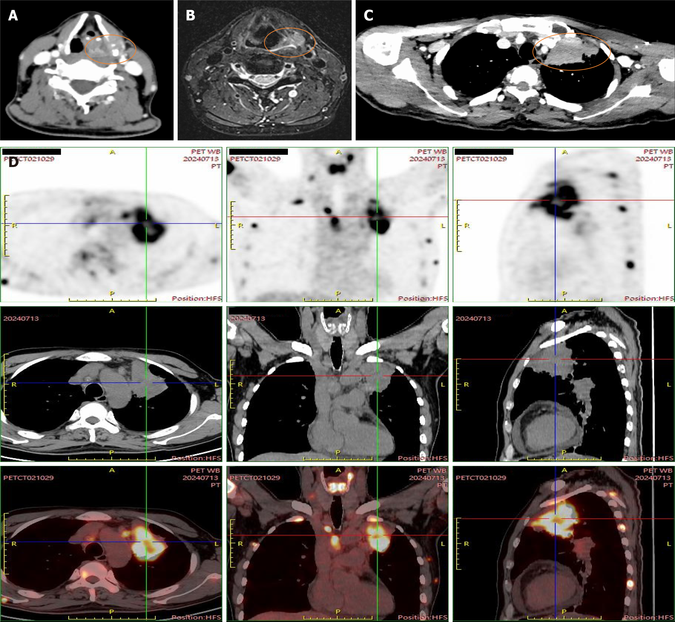
Figure 3 The imaging of the case.
A: Axial computed tomography (CT) with contrast image shows nodules in the left piriform sinus with bone destruction of the thyroid cartilage; B: Magnetic resonance imaging shows nodules in the left piriform sinus; C: CT with contrast image shows peripheral type lung cancer in the left upper lobe; D: Positron emission tomography-CT images. Imaging confirmed widespread metastases involving hilar/mediastinal/supraclavicular/cervical lymph nodes, bilateral adrenals, and multiple bones (spine and thyroid cartilage).
- Citation: Ai MM, Lin T, Guo RY, Zhang YY, Yu F. Unexpected metastasis of thyroid cartilage involvement from lung adenocarcinoma: A case report. World J Clin Cases 2025; 13(25): 107471
- URL: https://www.wjgnet.com/2307-8960/full/v13/i25/107471.htm
- DOI: https://dx.doi.org/10.12998/wjcc.v13.i25.107471


