©The Author(s) 2025.
World J Clin Cases. Sep 6, 2025; 13(25): 106089
Published online Sep 6, 2025. doi: 10.12998/wjcc.v13.i25.106089
Published online Sep 6, 2025. doi: 10.12998/wjcc.v13.i25.106089
Figure 1 Image of contrast-enhanced computed tomography.
A: Contrast-enhanced computed tomography (CE-CT) showing a retroperitoneal hematoma, indicating arterial bleeding; B: CE-CT showing celiac artery stenosis (arrow), supporting the diagnosis of median arcuate ligament syndrome; C: CE-CT showing two aneurysms in the inferior pancreaticoduodenal artery (arrow), requiring intervention.
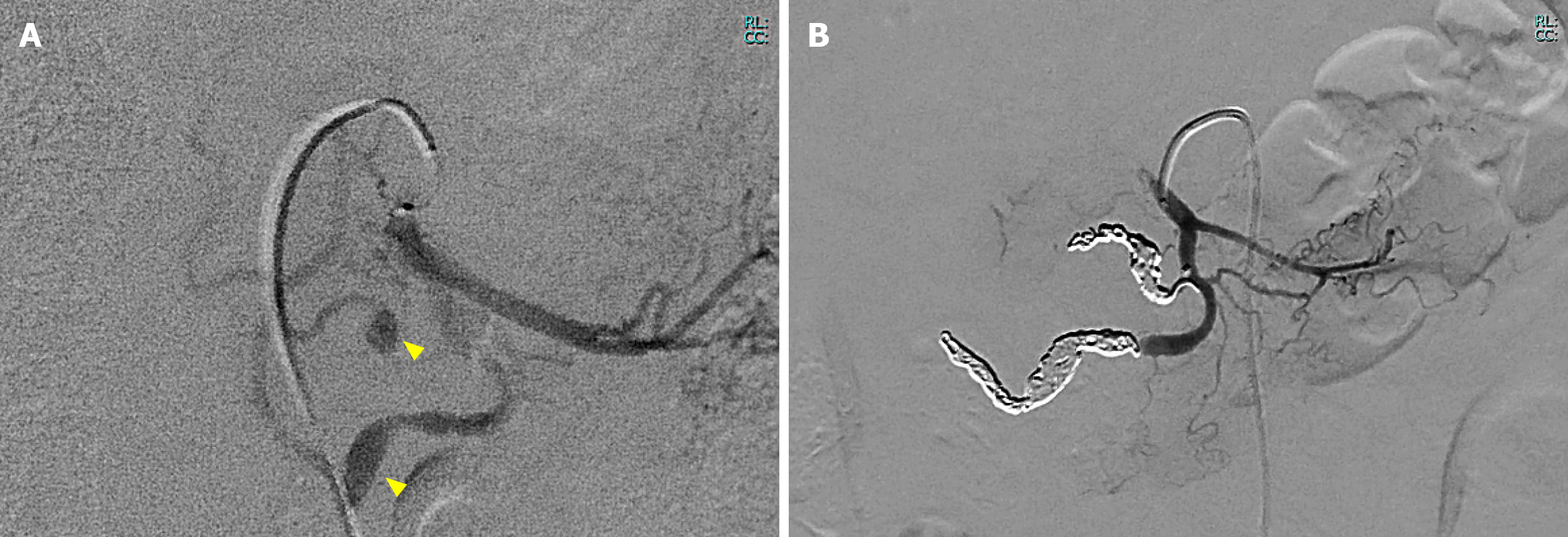
Figure 2 Images of angiogram during interventional radiology.
A: Angiography showing aneurysms in the inferior pancreaticoduodenal artery, confirming the bleeding source (arrow); B: Post-embolization angiography showing successful hemostasis with microcoils.
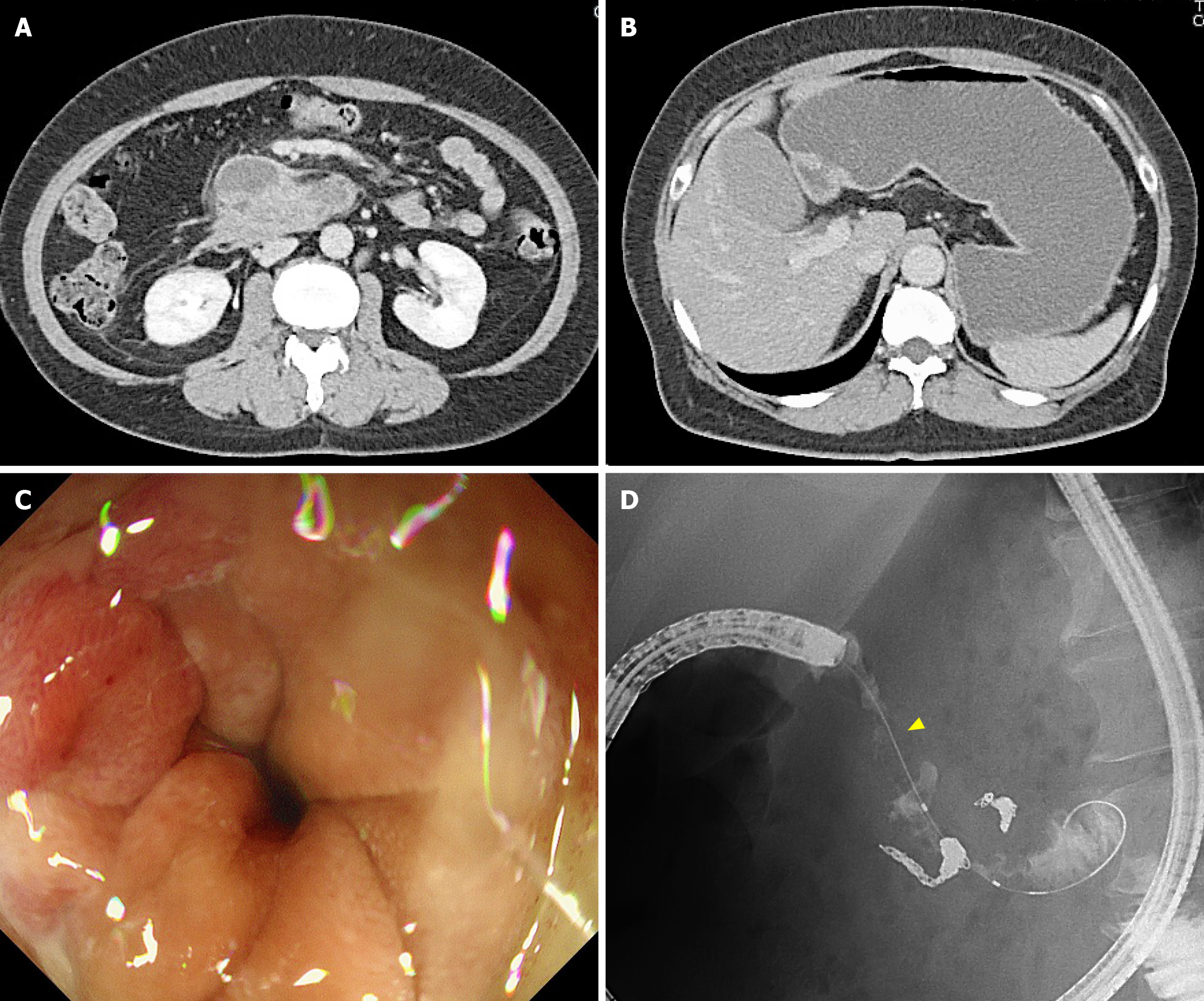
Figure 3 Duodenal stenosis following transcatheter arterial embolization on day 23.
A: Computed tomography (CT) showing gastric distension without duodenal dilation after transcatheter arterial embolization; B: CT showing reduced retroperitoneal hematoma; C: Endoscopy showing edematous duodenal mucosa without ulceration; D: Endoscopic contrast showing stenosis in the third part of the duodenum (arrow).
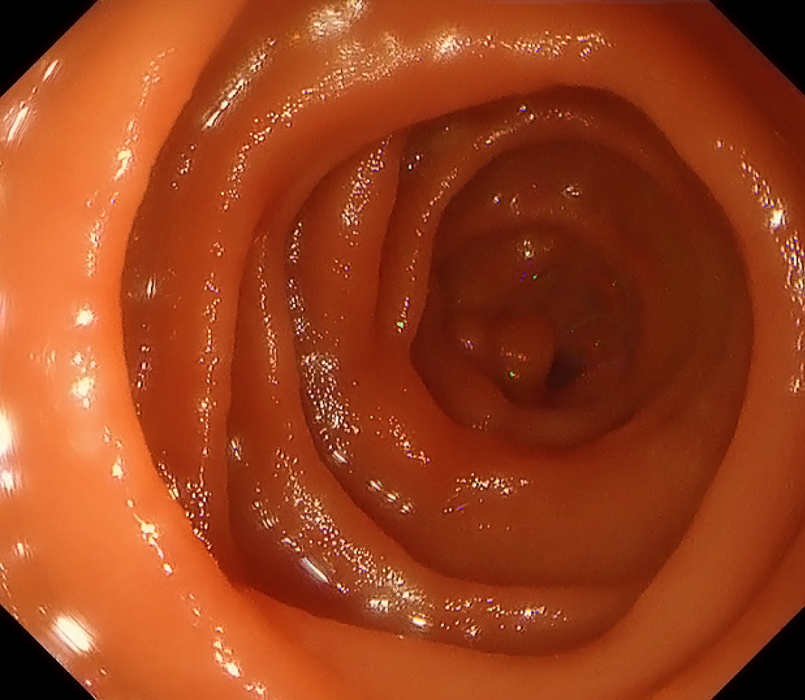
Figure 4 Endoscopy at 4 months showing resolved duodenal stenosis after surgery.
- Citation: Tanikawa T, Miyake K, Kawada M, Ishii K, Fushimi T, Urata N, Wada N, Nishino K, Suehiro M, Kawanaka M, Shiraha H, Haruma K, Fujiwara H, Yamatsuji T, Kawamoto H. Aneurysm rupture in median arcuate ligament syndrome leading to duodenal stenosis: A case report. World J Clin Cases 2025; 13(25): 106089
- URL: https://www.wjgnet.com/2307-8960/full/v13/i25/106089.htm
- DOI: https://dx.doi.org/10.12998/wjcc.v13.i25.106089


