©The Author(s) 2025.
World J Clin Cases. Sep 6, 2025; 13(25): 104134
Published online Sep 6, 2025. doi: 10.12998/wjcc.v13.i25.104134
Published online Sep 6, 2025. doi: 10.12998/wjcc.v13.i25.104134
Figure 1 The right eye had a corneal, self-sealed corneal scar, with siderotic pigments on the cataractous lens (blue arrow).
The right eye had a self-sealed capsular wound at the paracentral 5:00 position (white arrow), along with a corresponding iris defect beneath the corneal wound (yellow arrow).
Figure 2
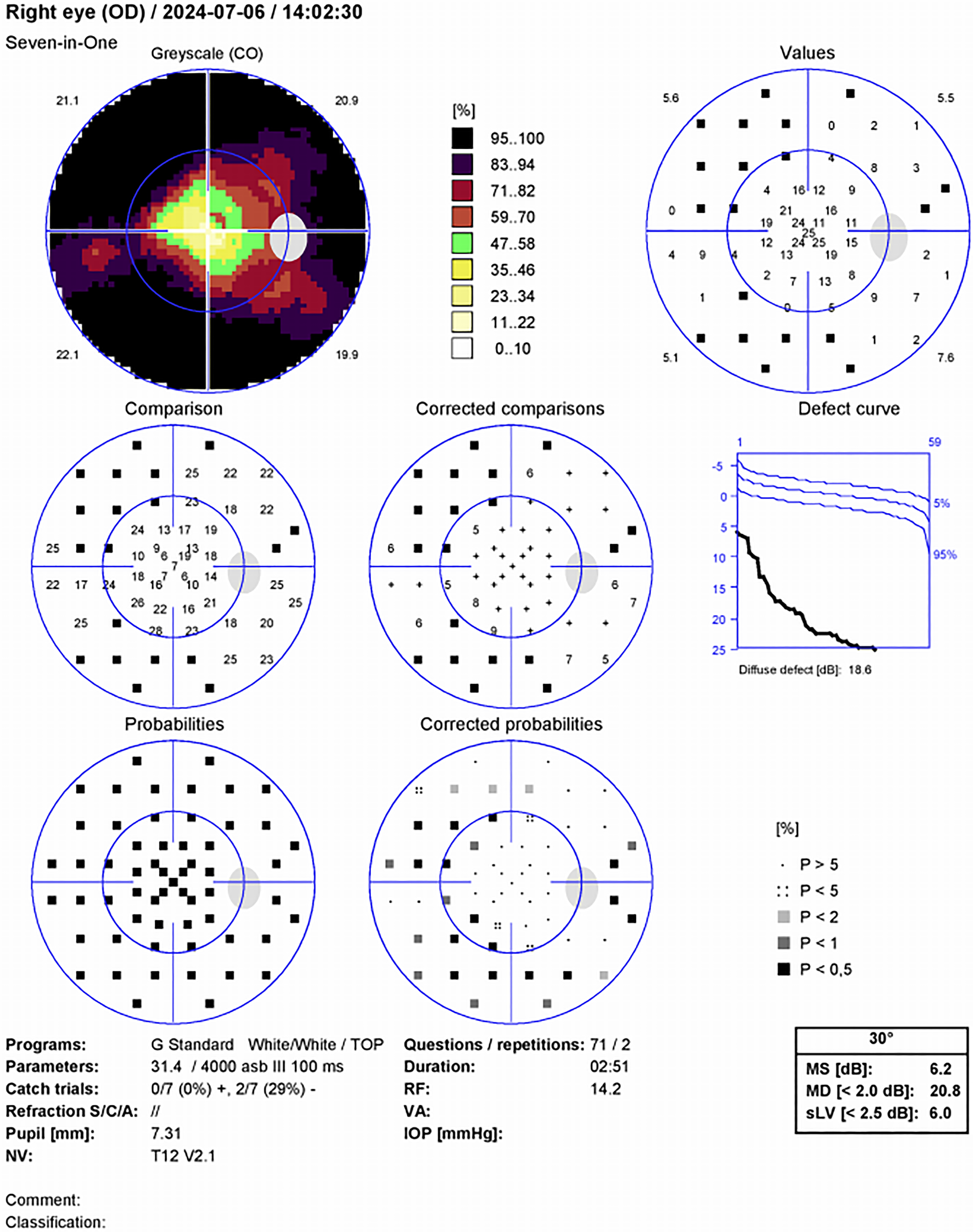
Visual field testing revealed tubular vision in the right eye.
Figure 3 No intraocular foreign bodies was detected on the B-scan.
Ultrasonic biomicroscopy image of the right eye. This image shows no detectable intraocular foreign bodies in the anterior chamber, iris stroma, ciliary body, or pars plana.
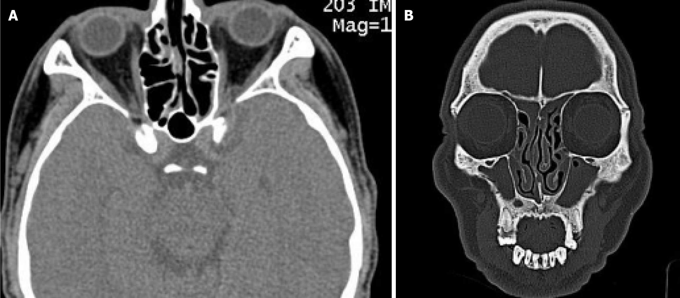
Figure 4 Coronal plain computed tomography of the orbit revealed no detectable intraocular radiopaque foreign bodies.
A: In both the axial views; B: In both the frontal views.
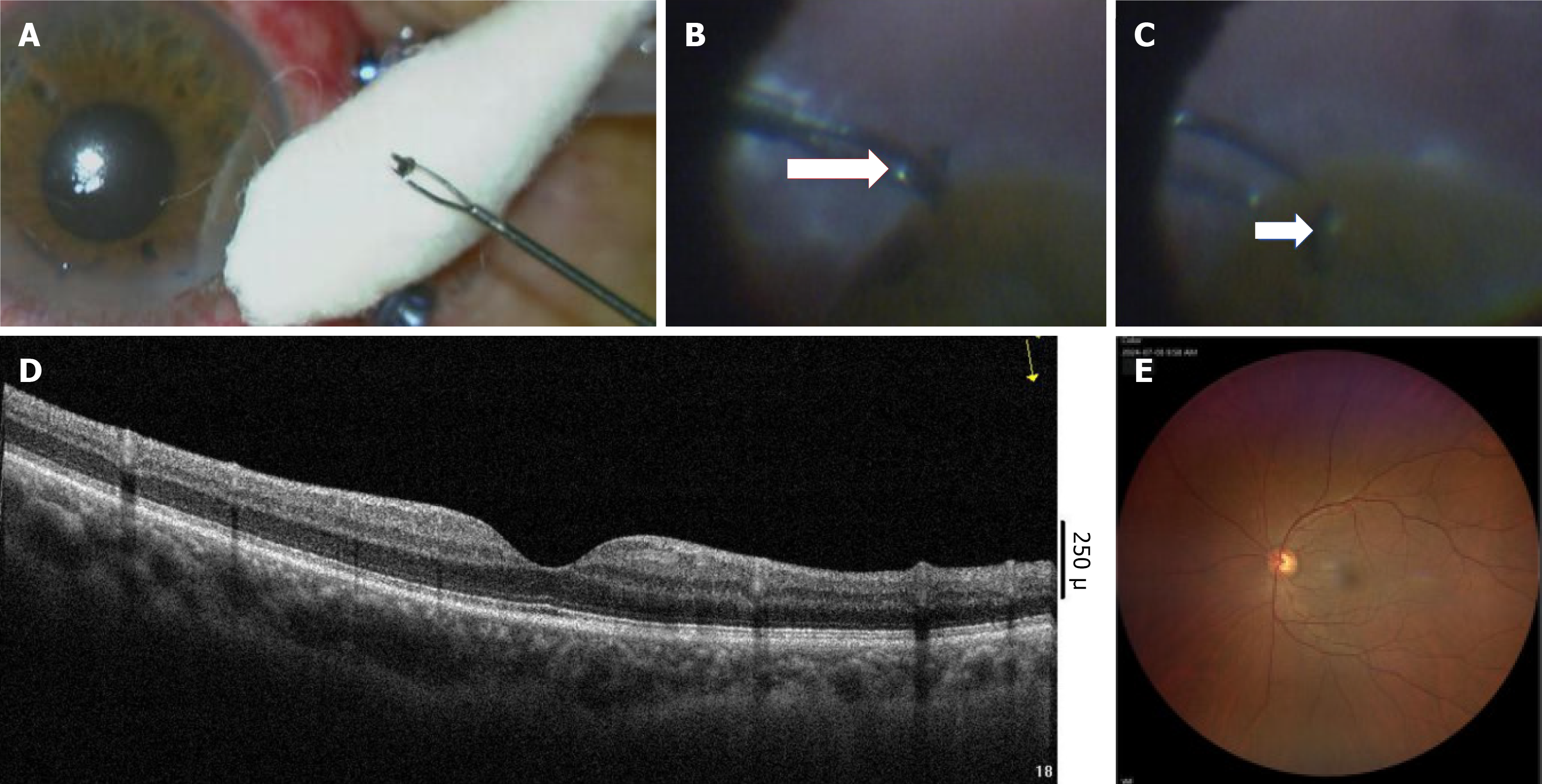
Figure 5 Inspection result.
A-C: The intraocular foreign body removed from the retina during vitreous surgery (the arrow in B indicates that the intraocular foreign body was located superior to the retina); D: Shows that the retinal and macular structures had recovered well 1 week after the operation; E: Showing that the fundus photography had recovered well 1 week after the operation.
- Citation: Xu LX, Kong YC. Ocular siderosis secondary to occult intraocular foreign body causing secondary glaucoma: A case report. World J Clin Cases 2025; 13(25): 104134
- URL: https://www.wjgnet.com/2307-8960/full/v13/i25/104134.htm
- DOI: https://dx.doi.org/10.12998/wjcc.v13.i25.104134


