Copyright
©The Author(s) 2025.
World J Clin Cases. Jul 16, 2025; 13(20): 105685
Published online Jul 16, 2025. doi: 10.12998/wjcc.v13.i20.105685
Published online Jul 16, 2025. doi: 10.12998/wjcc.v13.i20.105685
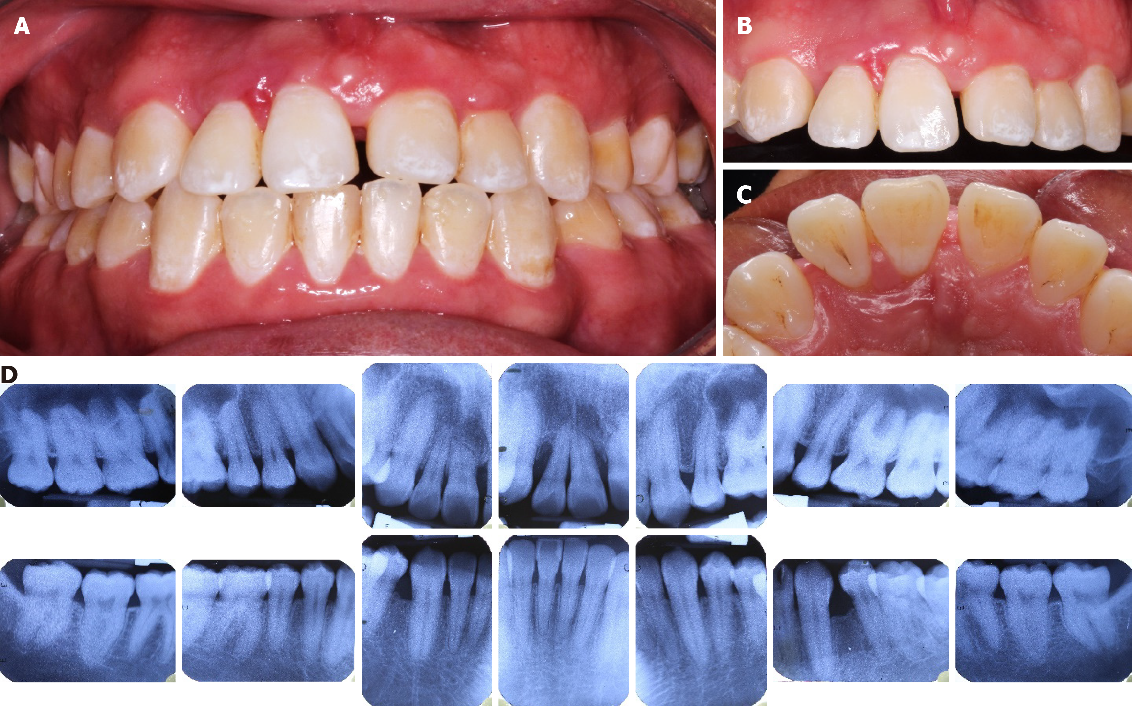
Figure 1 Initial intraoral photographs and radiographic.
A: Interoclusal view; B: Vestibular view; C: Palatal view; D: Periapical radiographic study.
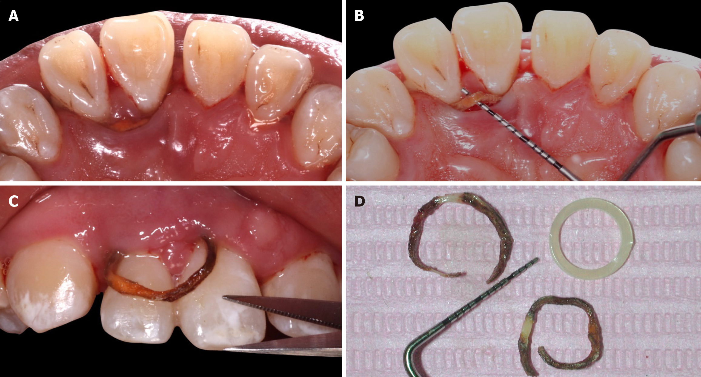
Figure 2 Elastic bands emerging from the gingival tissue during subgingival debridement.
A and B: Palatal views; C: Frontal view; D: Comparison of the new orthodontic elastic with the two elastics removed.
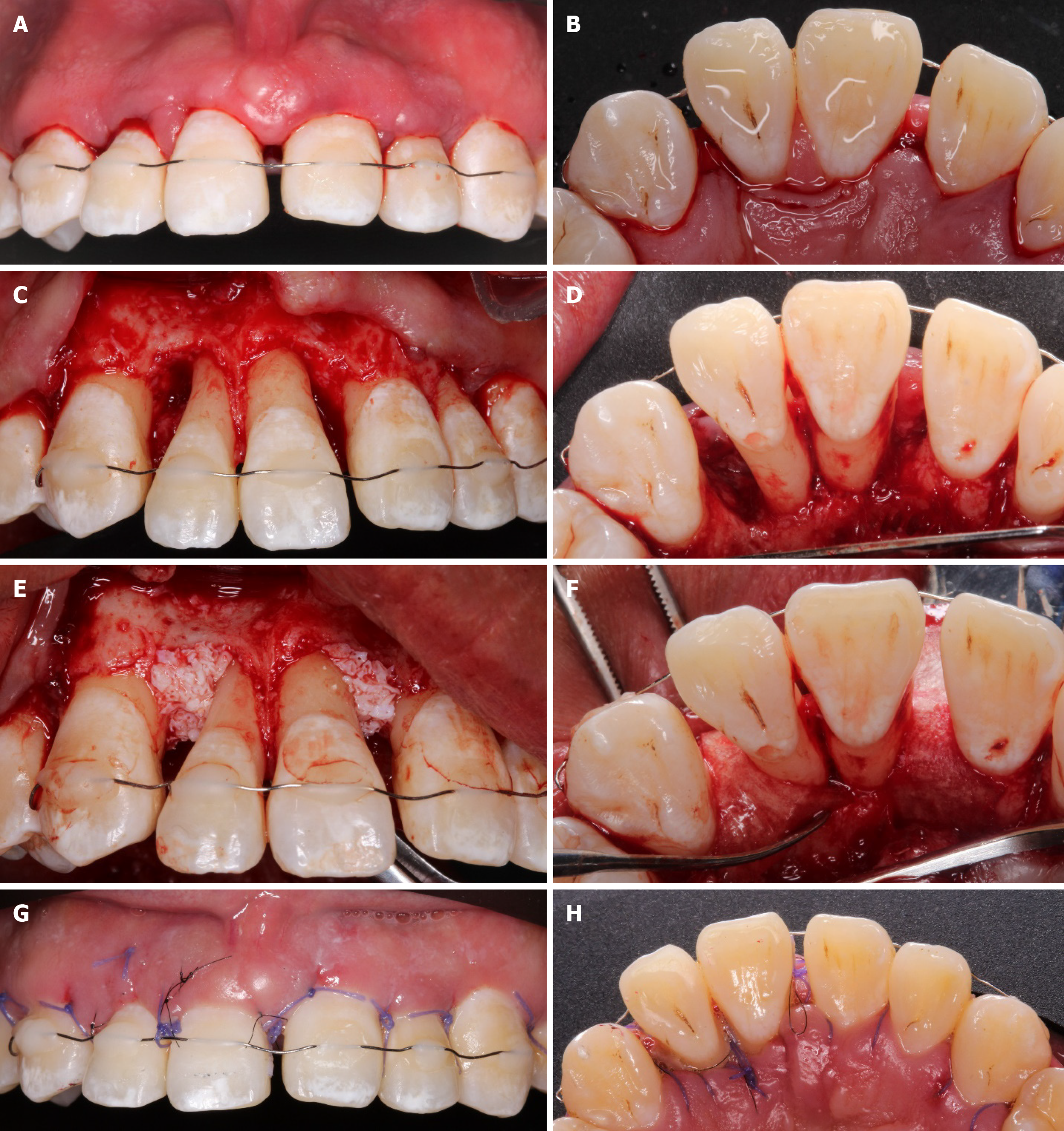
Figure 3 Surgical procedure.
A: Intrasurcular incision of the teeth 14 to 23; B: Incisions to preserve papillae on the palatal surface in teeth 11, 12, 13, and 21; C and D: Combined bone defects; E: Bovine bone placement; F: Placement of resorbable collagen membrane; G and H: Simple and suspensory stitches.
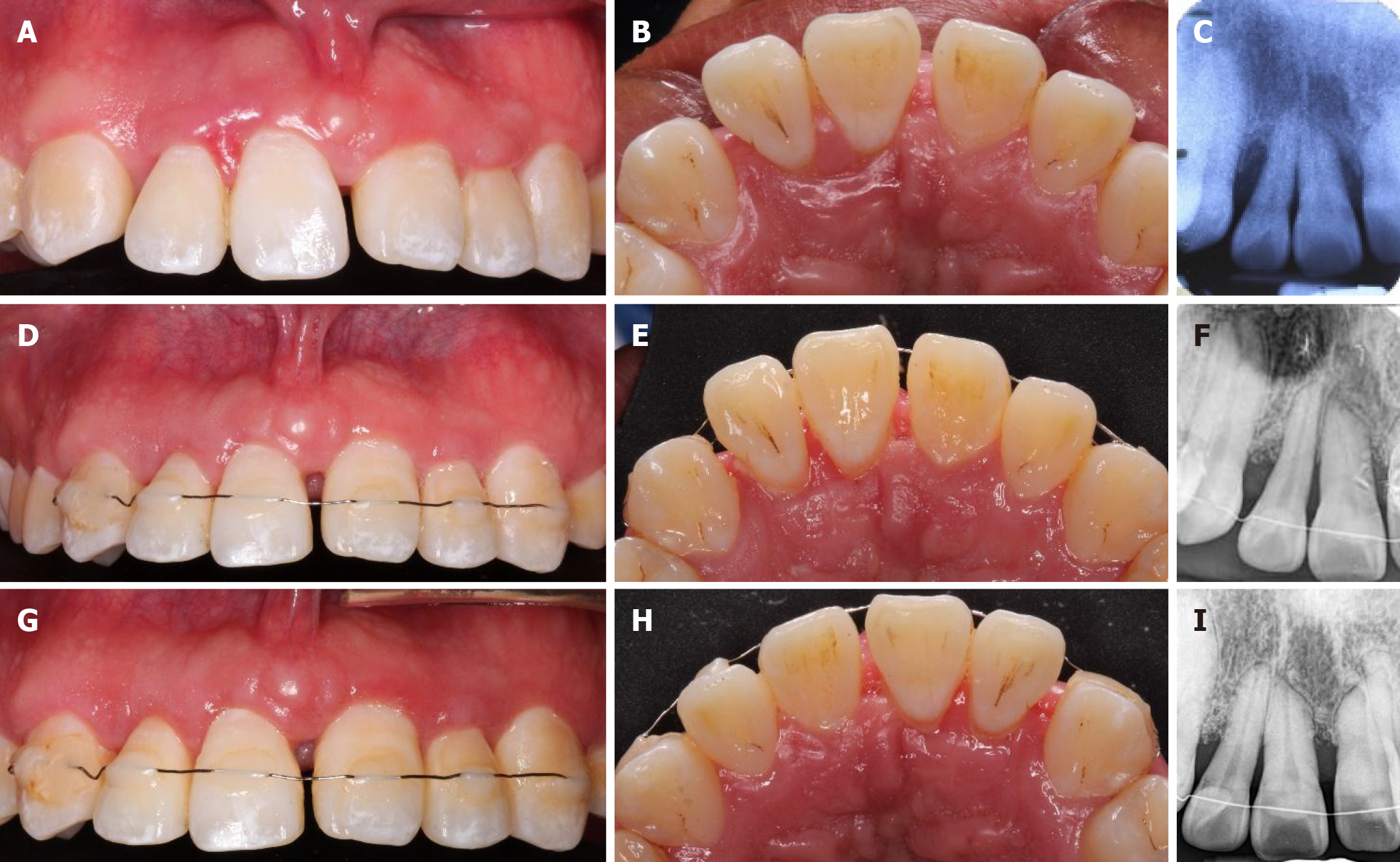
Figure 4 Case follow-up.
A and B: Initial images; C: Initial radiograph; D and E: Clinical images 6 months after regenerative surgery; F: X-ray 6 months after surgery; G and H: 2 years post-surgical; I: X-ray 2 years after surgery.
- Citation: Montiel-López PA, García-Nuñez JC, Muro-Jiménez ML, Soto-Chávez AA, Martínez-Rodríguez VM, Rodríguez-Montaño R, Ruiz-Gutiérrez AC. Management of intrabony defects associated with the iatrogenic use of orthodontic elastic bands: A case report. World J Clin Cases 2025; 13(20): 105685
- URL: https://www.wjgnet.com/2307-8960/full/v13/i20/105685.htm
- DOI: https://dx.doi.org/10.12998/wjcc.v13.i20.105685


