©The Author(s) 2025.
World J Clin Cases. Jul 6, 2025; 13(19): 104400
Published online Jul 6, 2025. doi: 10.12998/wjcc.v13.i19.104400
Published online Jul 6, 2025. doi: 10.12998/wjcc.v13.i19.104400
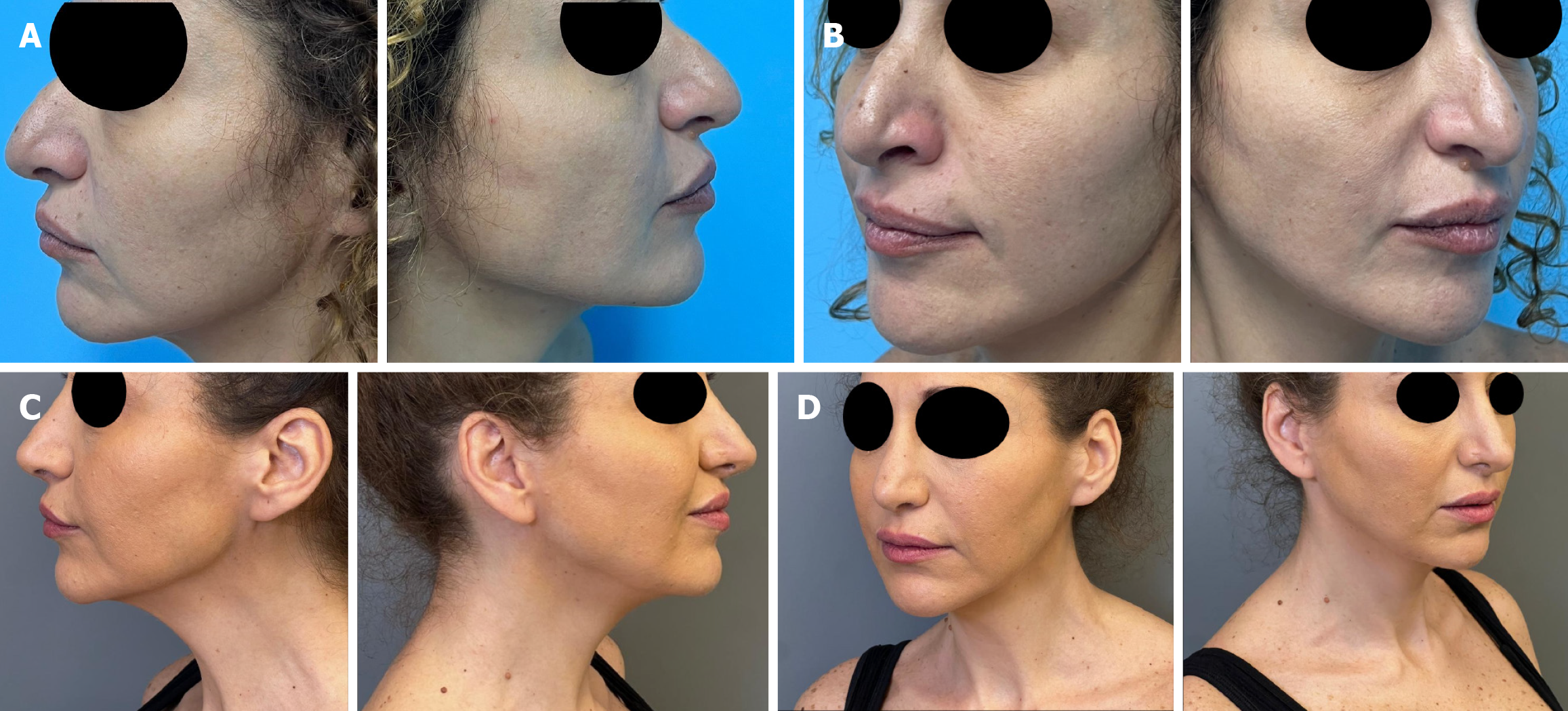
Figure 1 View photos of the patient before and after surgery.
A: Pre-operative side view photos of the patient; B: Post-operative (6 months) side view photos of the patient; C: Pre-operative three-quarter view photos of the patient; D: Post-operative (6 months) three-quarter view photos of the patient.
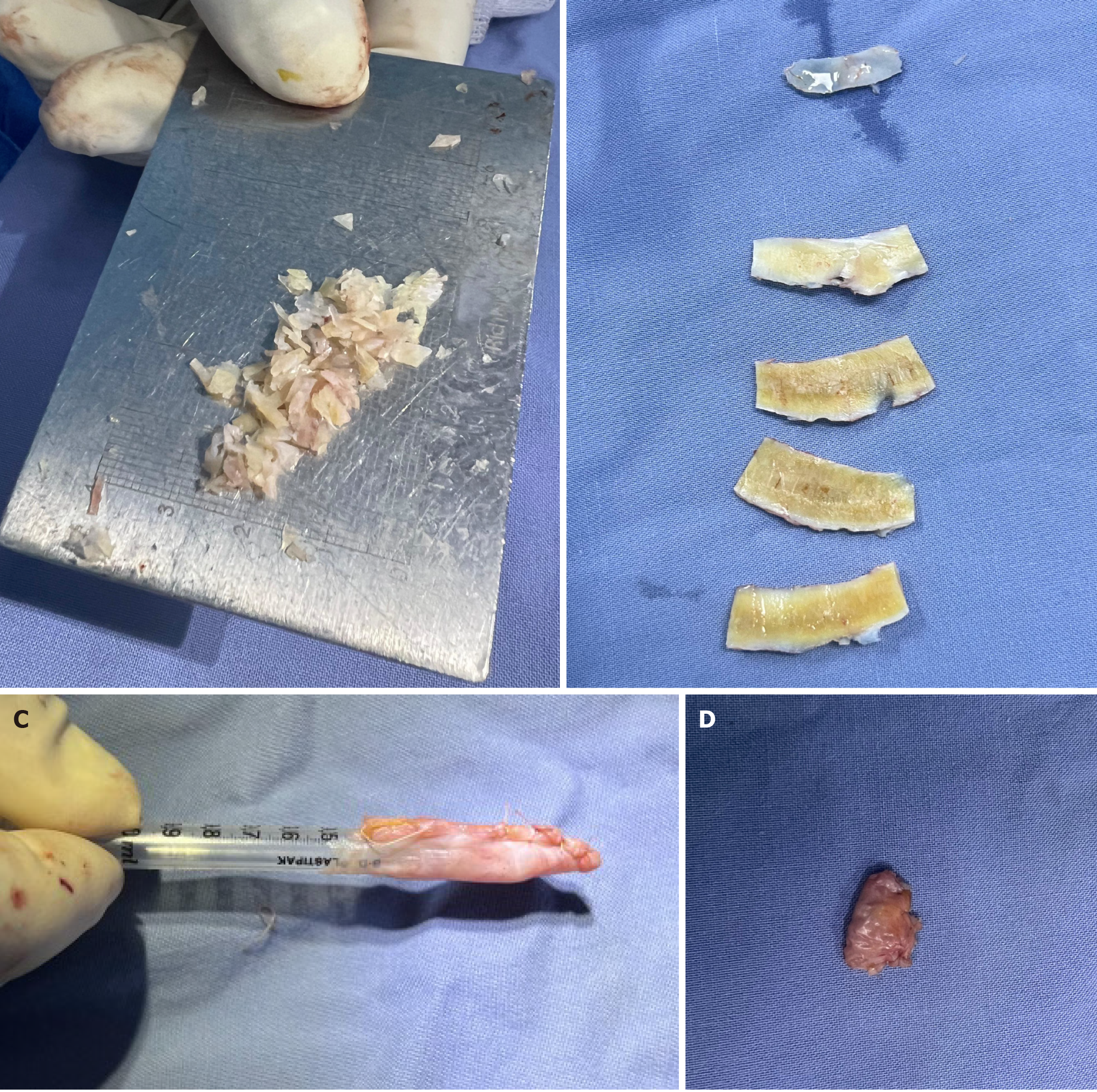
Figure 2 Preparation of cartilage and capsule.
A: Diced cartilage; B: Rib cartilage sculpted in lamellaes; C: Part of capsule prepared; D: Capsule wrapping diced cartilage.
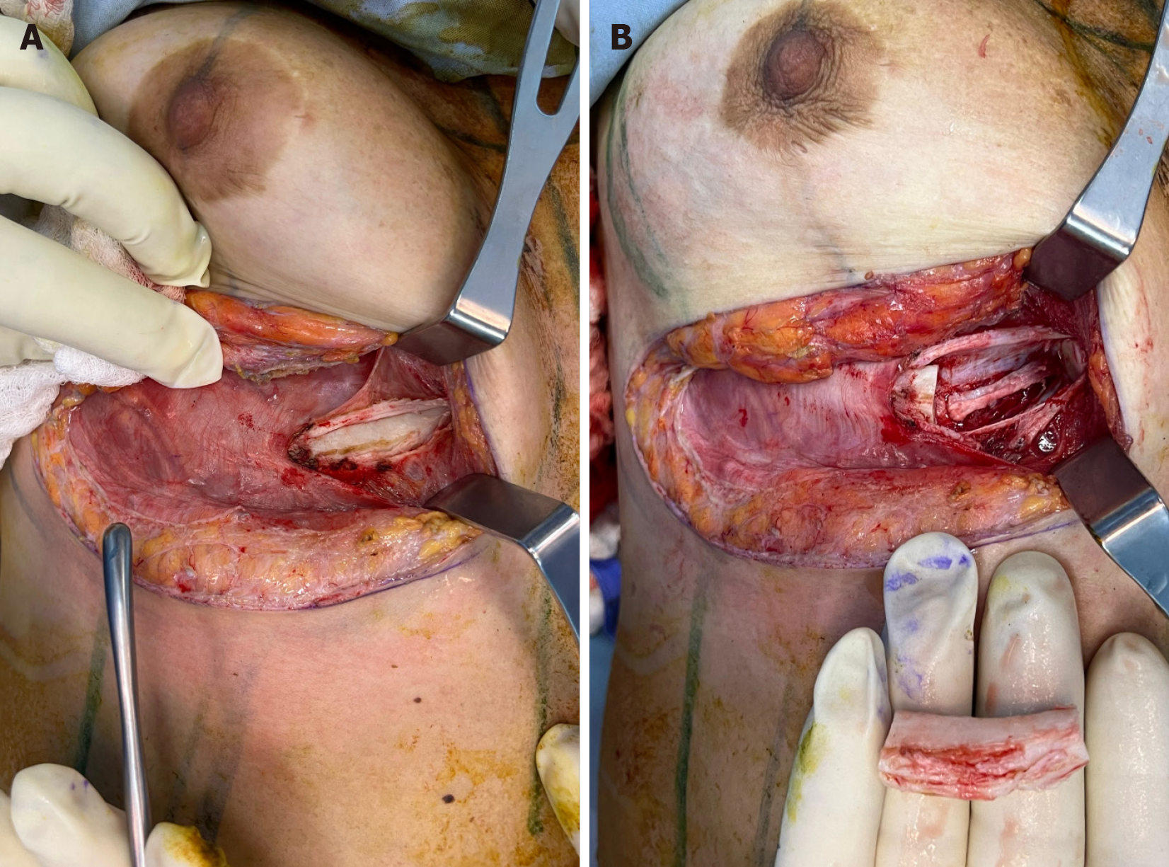
Figure 3 Approach to the fifth rib cartilage via inframammary fold incision.
A: Before removal of right 5th rib cartilage; B: After removal of right 5th rib cartilage.
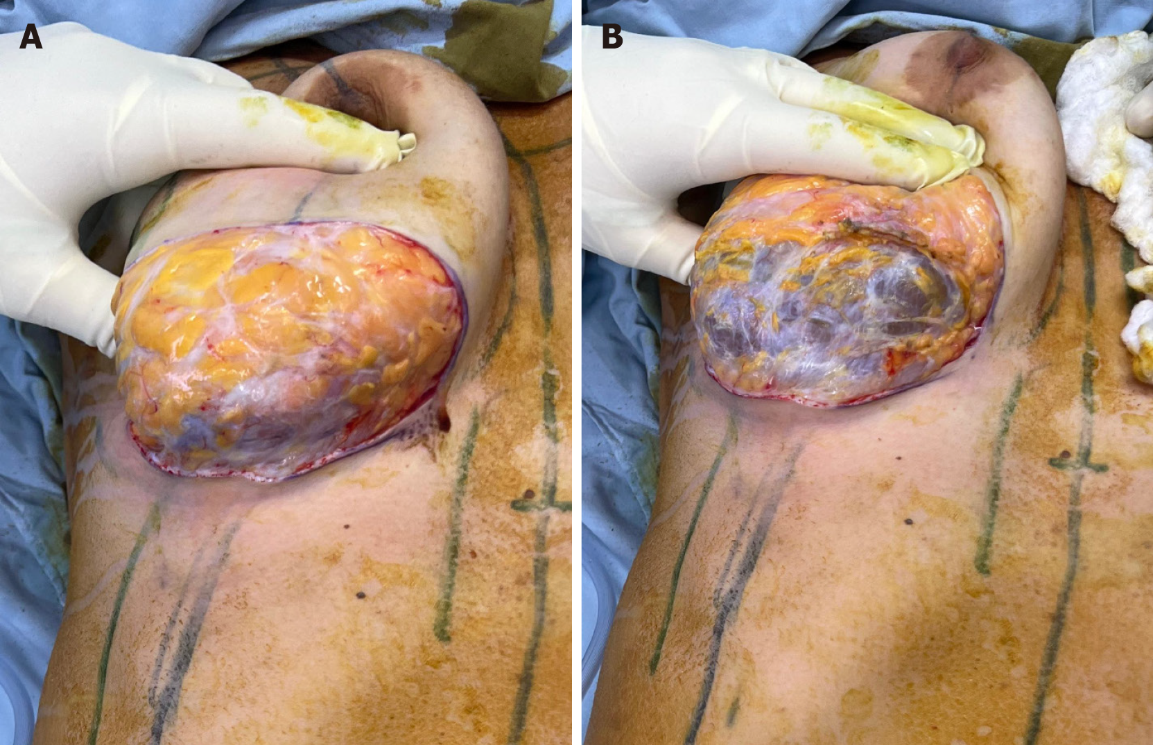
Figure 4 Exposure of breast implant capsule.
A: Exposure of the breast implant capsule with surrounding fibrotic tissue; B: Removal of the breast implant capsule, revealing its internal structure.
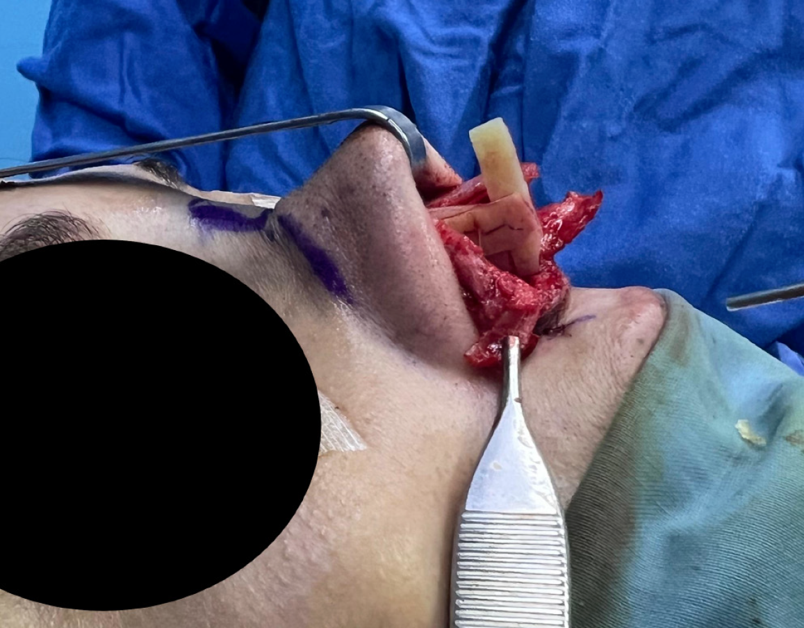
Figure 5
Columellar strut fixed to the extended spreader grafts.
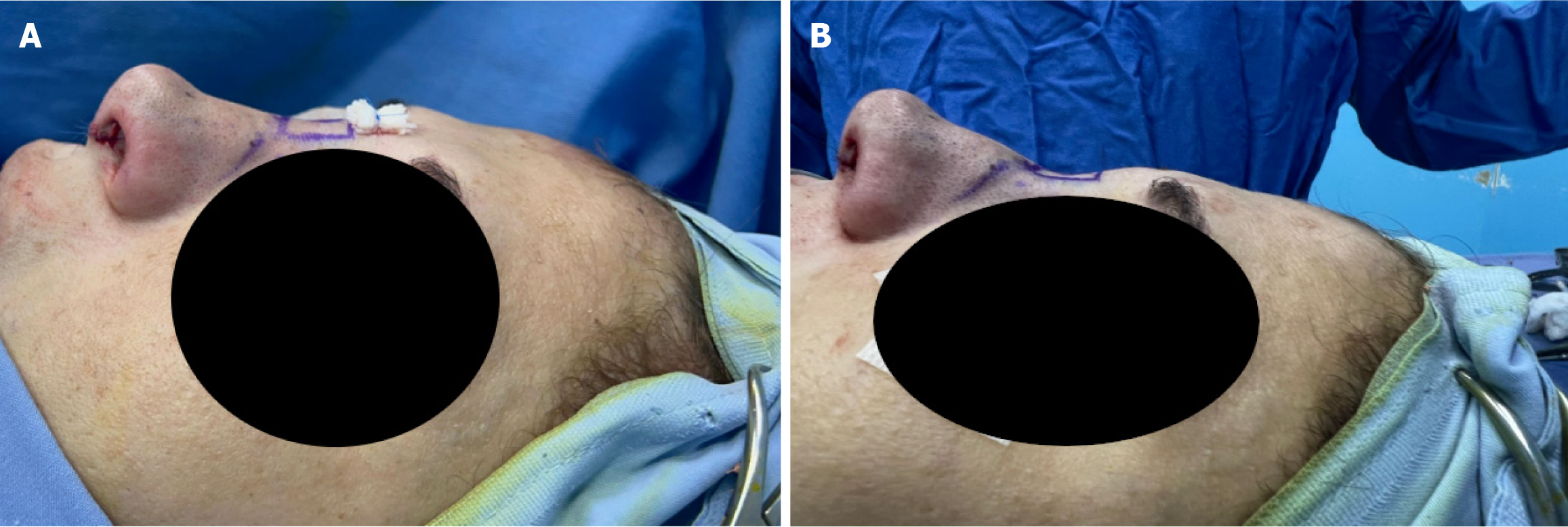
Figure 6 Before and after introducing capsular-wrapped diced cartilage.
A: After introducing the capsular-wrapped diced cartilage; B: Before introducing capsular-wrapped diced cartilage.
- Citation: Stephan H, Rihani H, Dagher E, El Choueiri J. Diced cartilage in capsula based on diced cartilage in fascia technique: A case report. World J Clin Cases 2025; 13(19): 104400
- URL: https://www.wjgnet.com/2307-8960/full/v13/i19/104400.htm
- DOI: https://dx.doi.org/10.12998/wjcc.v13.i19.104400


