Copyright
©The Author(s) 2025.
World J Clin Cases. Apr 26, 2025; 13(12): 99229
Published online Apr 26, 2025. doi: 10.12998/wjcc.v13.i12.99229
Published online Apr 26, 2025. doi: 10.12998/wjcc.v13.i12.99229
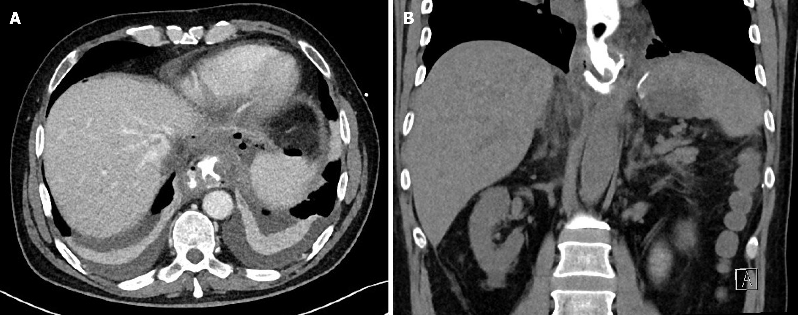
Figure 1 Computed tomography scan with oral contrast demonstrating oral contrast filling a fluid collection to the left of the esopha
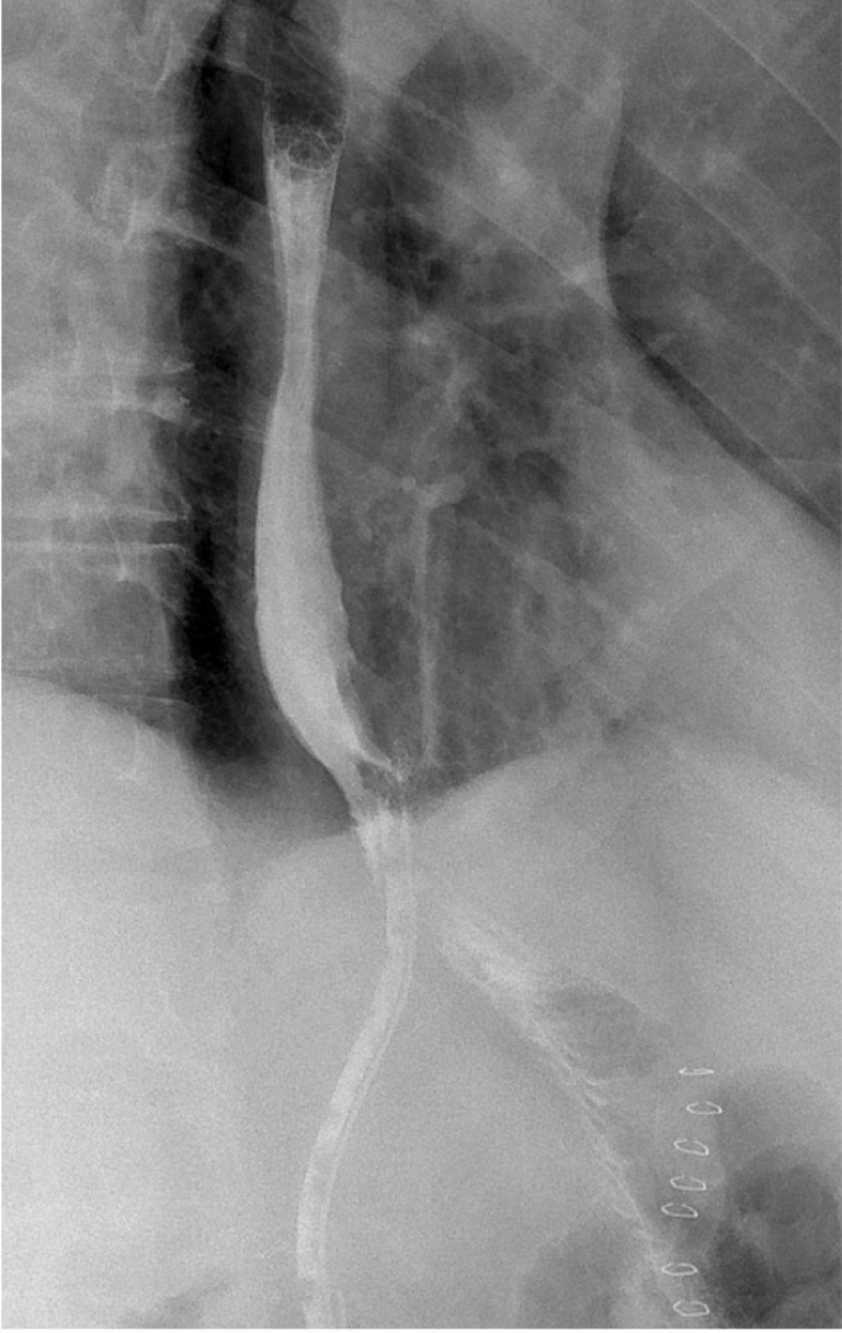
Figure 2 Contrast swallow radiograph demonstrating contrast filling of the abdominal drain without evidence of extraluminal extra
Figure 3 Endoscopy on postoperative day 59 revealed iatrogenic perforation of the jejunum secondary to drain migration.
The perforation site was located approximately 2 cm distal to the intact esophagojejunostomy (EJA), which showed no signs of dehiscence. A: The tip of the drain within the esophagus; B: The perforation site near the EJA; C: An intact EJA with surgical clips visible.
- Citation: Janež J, Romih J, Čebron Ž, Gavric A, Plut S, Grosek J. Intraluminal migration of a surgical drain near an anastomosis site after total gastrectomy: A case report. World J Clin Cases 2025; 13(12): 99229
- URL: https://www.wjgnet.com/2307-8960/full/v13/i12/99229.htm
- DOI: https://dx.doi.org/10.12998/wjcc.v13.i12.99229


