Copyright
©The Author(s) 2025.
World J Clin Cases. Apr 26, 2025; 13(12): 100045
Published online Apr 26, 2025. doi: 10.12998/wjcc.v13.i12.100045
Published online Apr 26, 2025. doi: 10.12998/wjcc.v13.i12.100045
Figure 1 Oral small bowel imaging.
A-C: Visible elevation of the small intestine and bowel occupation with hemorrhagic manifestations.
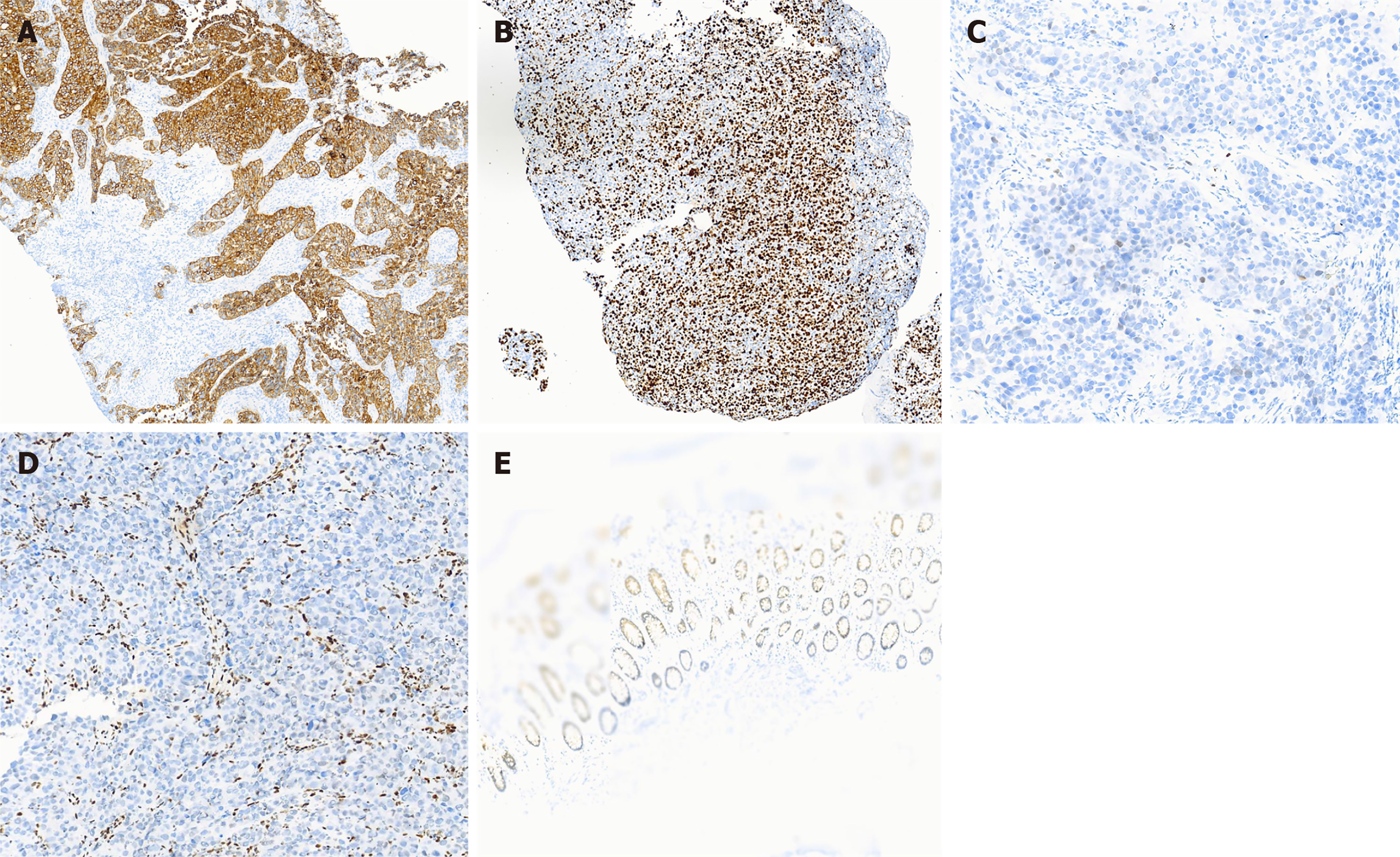
Figure 2 Immunohistochemistry results.
A: CK7 is a key component of cytokeratin, and the positivity is usually indicative of a high likelihood of adenocarcinoma. In this patient, CK7 was distinctly positive in a localized area; B: The primary site of Ki67 positivity is located in the nucleus. Ki67 is a marker of cell proliferation and primarily reflects the proliferative state of the cell. The immunohistochemistry result of the small bowel microscopy pathology of this patient shows a Ki67 index of 65%, reflecting a high proliferative activity). CA weakly positive expression of SATB2 is observed in the individual; D: SMARCA4-deficient undifferentiated carcinomas are commonly found in multiple sites, most especially in the lungs and rarely in the gastrointestinal tract. Based on a 2023 lung tissue biopsy that also demonstrated SMARCA4 deletion, intestinal metastases from lung cancer cannot be excluded from this intestinal tissue; E: Weak, localized positivity for Villin was observed.
- Citation: Yuan TY, Chen YX, Zhao YG, Wang B, Wang SX. Gastrointestinal bleeding due to small bowel metastasis from lung adenocarcinoma: A case report. World J Clin Cases 2025; 13(12): 100045
- URL: https://www.wjgnet.com/2307-8960/full/v13/i12/100045.htm
- DOI: https://dx.doi.org/10.12998/wjcc.v13.i12.100045


