Copyright
©The Author(s) 2024.
World J Clin Cases. Mar 6, 2024; 12(7): 1313-1319
Published online Mar 6, 2024. doi: 10.12998/wjcc.v12.i7.1313
Published online Mar 6, 2024. doi: 10.12998/wjcc.v12.i7.1313
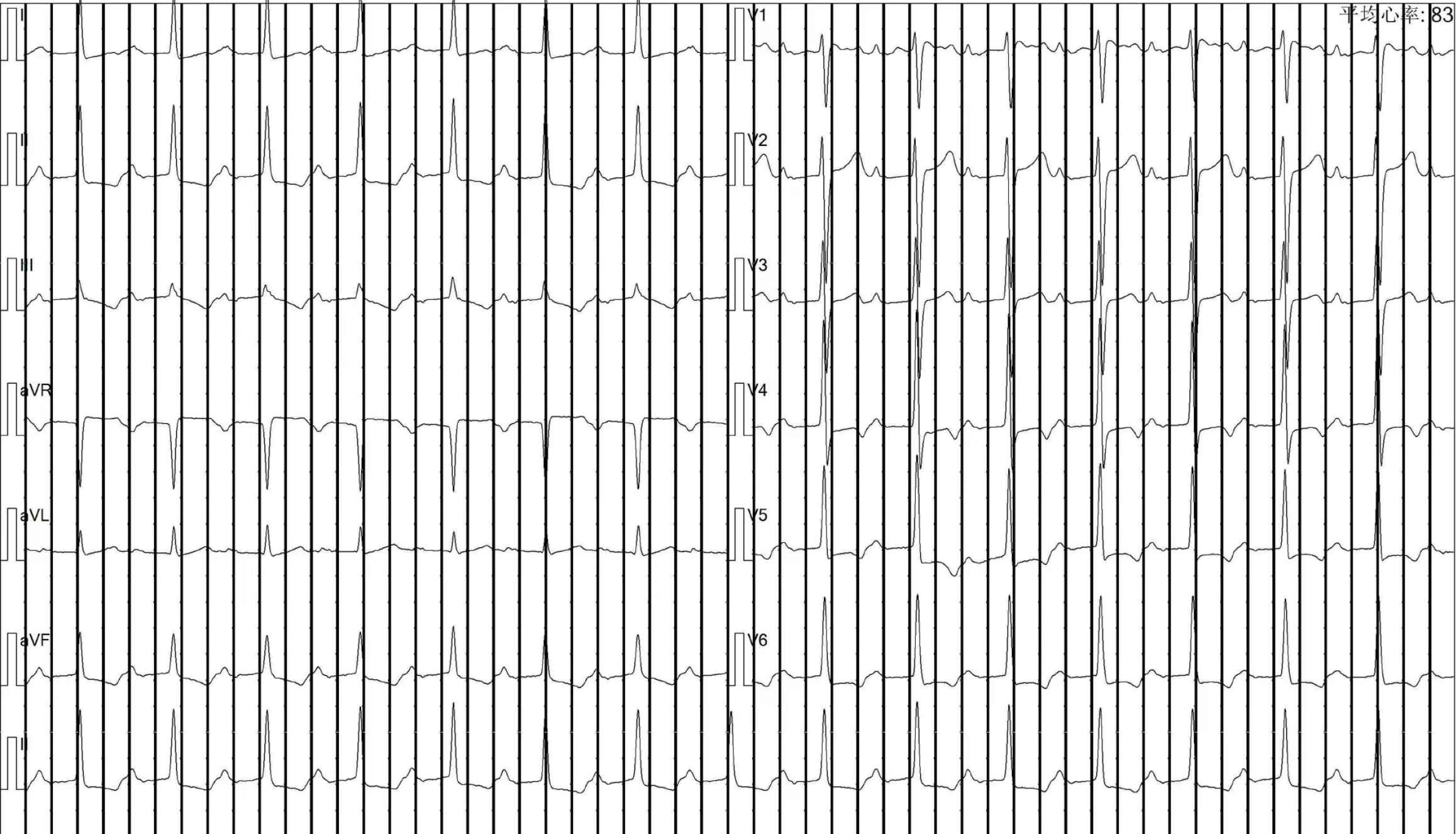
Figure 1 Her electrocardiograph from 2018 showed a first-degree atrioventricular block.
The PR-interval was 310 ms.
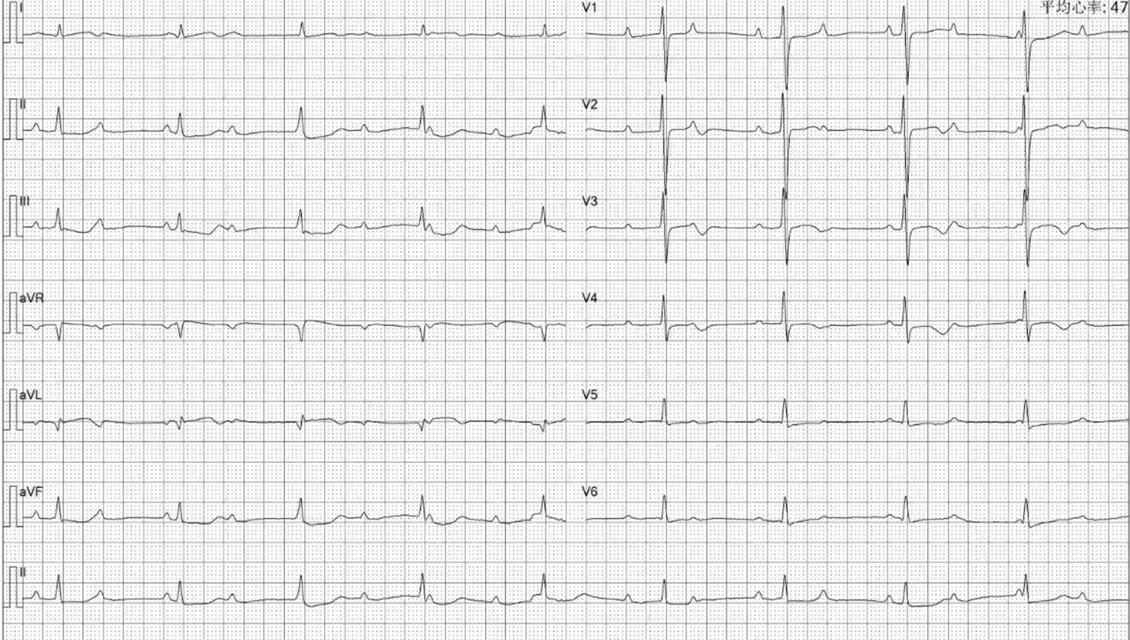
Figure 2 Her electrocardiograph in 2020 showed a complete atrioventricular block.
Figure 3 Comparison of Echocardiography data from 2021 and 2022.
A and B: Color Doppler echocardiography showing calcification in the posterior leaflet of the mitral valve. The calcification point in the posterior mitral lobe was lower in A than in B. A was obtained beginning in 2021, and B was obtained beginning in 2022. Arrow: The calcified area of the mitral valve.
Figure 4 Comparison of chest computed tomography data from 2021 (B and D) and 2022 (A and C).
Chest computed tomography revealed that the calcifications of the left atrium and left ventricle improved.
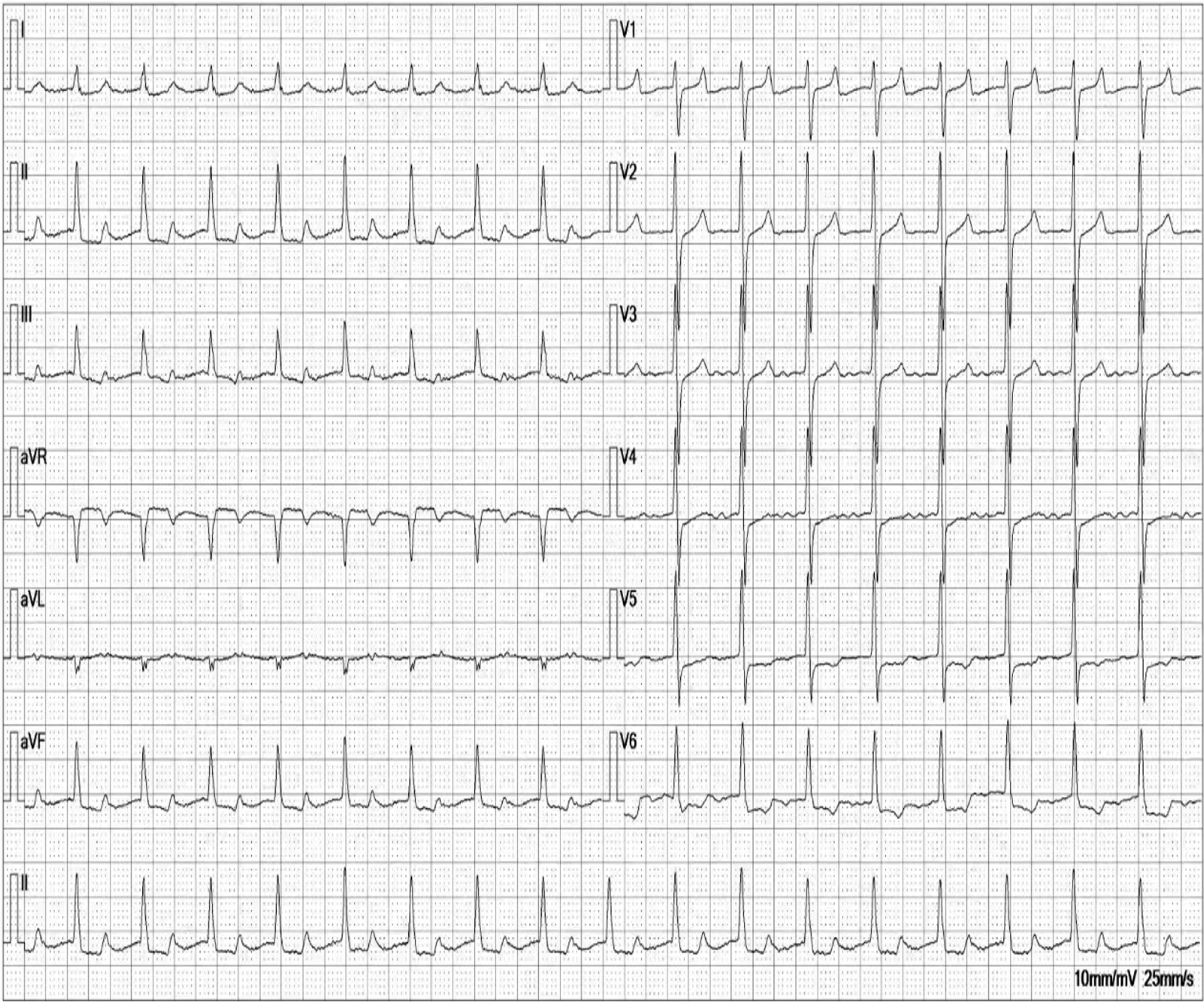
Figure 5 An electrocardiograph from 2022 showing a return to a first-degree atrioventricular block.
The PR-interval was 342 ms.
- Citation: Xu SS, Hao LH, Guan YM. Reversal of complete atrioventricular block in dialysis patients following parathyroidectomy: A case report. World J Clin Cases 2024; 12(7): 1313-1319
- URL: https://www.wjgnet.com/2307-8960/full/v12/i7/1313.htm
- DOI: https://dx.doi.org/10.12998/wjcc.v12.i7.1313


