Copyright
©The Author(s) 2024.
World J Clin Cases. Dec 6, 2024; 12(34): 6728-6735
Published online Dec 6, 2024. doi: 10.12998/wjcc.v12.i34.6728
Published online Dec 6, 2024. doi: 10.12998/wjcc.v12.i34.6728
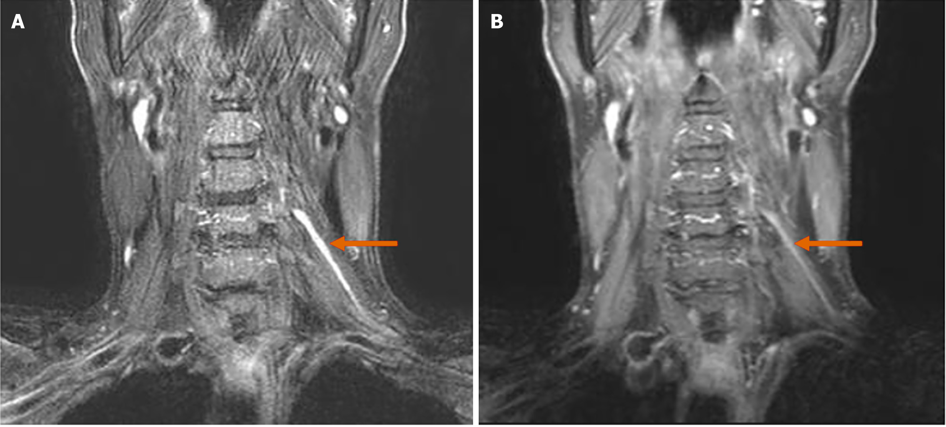
Figure 1 Brachial plexus magnetic resonance imaging.
A: T2-weighted fat saturated coronal images; B: T1-weighted fat saturated contrast enhanced coronal image. Asymmetric thickening and increased signal intensity with mild contrast enhancement of left C5 nerve root/trunk level is noted (arrows).
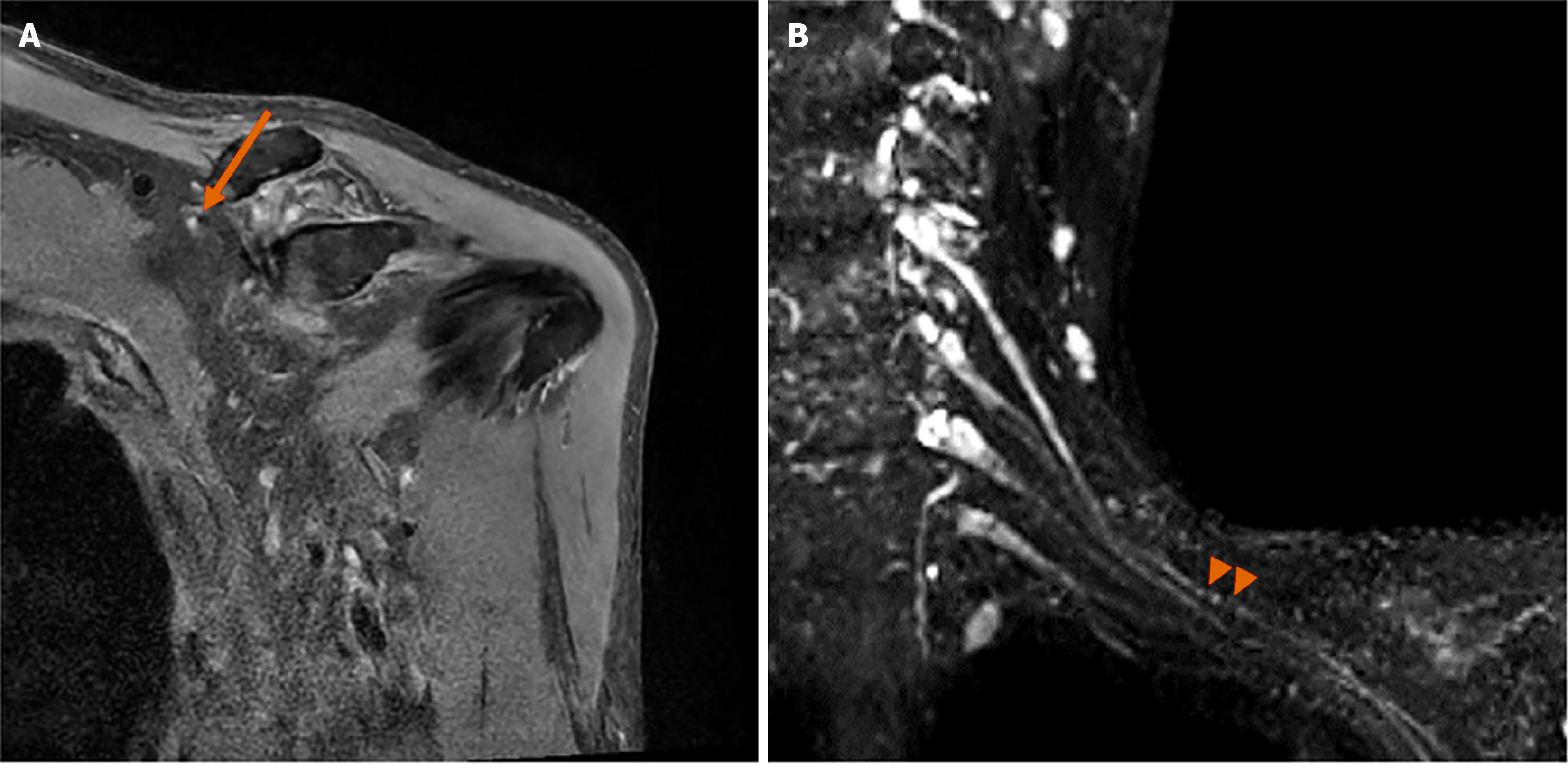
Figure 2 Intermediated-weighted fat saturated oblique coronal image.
A: Shoulder magnetic resonance imaging; B: Magnetic resonance (MR) neurography of brachial plexus. Focal thickening and increased signal intensity of left suprascapular nerve is noted (arrow). MR neurography demonstrates focal constriction of left suprascapular nerve (arrow heads).
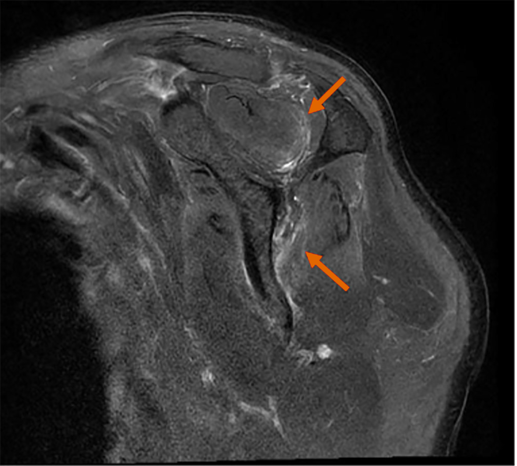
Figure 3 T2-weighted fat saturated oblique sagittal image of shoulder magnetic resonance imaging.
Increased T2 signal intensities of both supraspinatus (upper arrow) and infraspinatus (lower arrow) muscles are noted.
- Citation: Bang MH, Song HL, Hahn S, Kim W, Do HK. Neuralgic amyotrophy with hourglass-like constrictions: A case report. World J Clin Cases 2024; 12(34): 6728-6735
- URL: https://www.wjgnet.com/2307-8960/full/v12/i34/6728.htm
- DOI: https://dx.doi.org/10.12998/wjcc.v12.i34.6728


