Copyright
©The Author(s) 2024.
World J Clin Cases. Dec 6, 2024; 12(34): 6687-6695
Published online Dec 6, 2024. doi: 10.12998/wjcc.v12.i34.6687
Published online Dec 6, 2024. doi: 10.12998/wjcc.v12.i34.6687
Figure 1 Workflow of the study.
PACS: Picture archiving and communication system; MR: Magnetic resonance; mSBA: Modified skull base angle; SBA: Skull base angle.
Figure 2 Measurement technique of skull base angles.
A and C: The angle formed between the line drawn from the nasion to the center of the hypophysis gland and another line from this point to the anterior border of the foramen magnum was measured as the skull base angle (SBA); B and D: The line from the tip of the dorsum sella extending across the anterior cranial fossa and another line drawn along the posterior margin of the clivus forms the modified SBA.
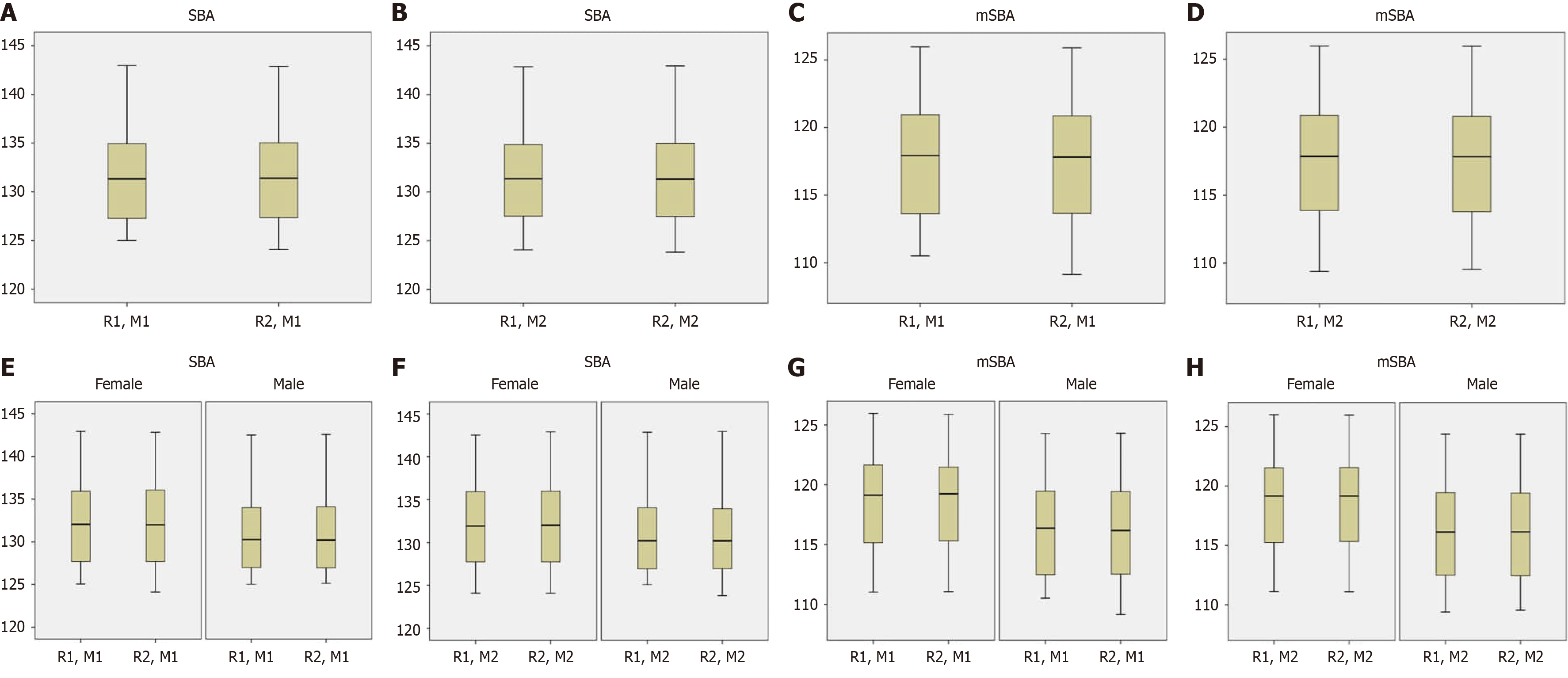
Figure 3 Box-plot analyses showing the data distributions of skull base angle and modified skull base angle.
A, B, E and F: Skull base angle (SBA); C, D, G and H: Modified SBA (mSBA); E, F, G and H: The comparison of data distributions between female and male groups. Similar data distributions were obtained between the reviewers. In addition, lower SBA and mSBA values were observed for the male group in comparison with the females as shown by the box-plots. M1: First measurements; M2: Second measurements; R1: Reviewer 1; R2: Reviewer 2.
- Citation: Kizilgoz V, Aydin S, Aydemir H, Keles P, Kantarci M. Interobserver and intraobserver reliability of skull base angles measured on magnetic resonance images. World J Clin Cases 2024; 12(34): 6687-6695
- URL: https://www.wjgnet.com/2307-8960/full/v12/i34/6687.htm
- DOI: https://dx.doi.org/10.12998/wjcc.v12.i34.6687


