Copyright
©The Author(s) 2024.
World J Clin Cases. Oct 16, 2024; 12(29): 6302-6306
Published online Oct 16, 2024. doi: 10.12998/wjcc.v12.i29.6302
Published online Oct 16, 2024. doi: 10.12998/wjcc.v12.i29.6302
Figure 1
Abdominal ultrasound demonstrating swollen head of the pancreas with the suggestion of peripancreatic fluid.
Figure 2 Abdominal computed tomography: Pancreas is bulky showing inhomogeneous enhancement, with patchy non-enhancing areas.
Internal calcifications are seen. Minimal surrounding inflammatory changes are also noted. The pancreatic duct is dilated.
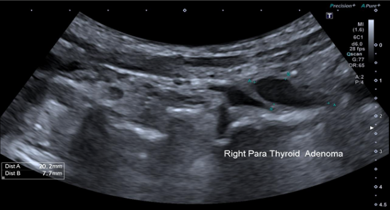
Figure 3
Ultrasound of the neck showed a large parathyroid adenoma at the inferior lobe pole of the right lobe of the thyroid medially, measuring 207 mm.
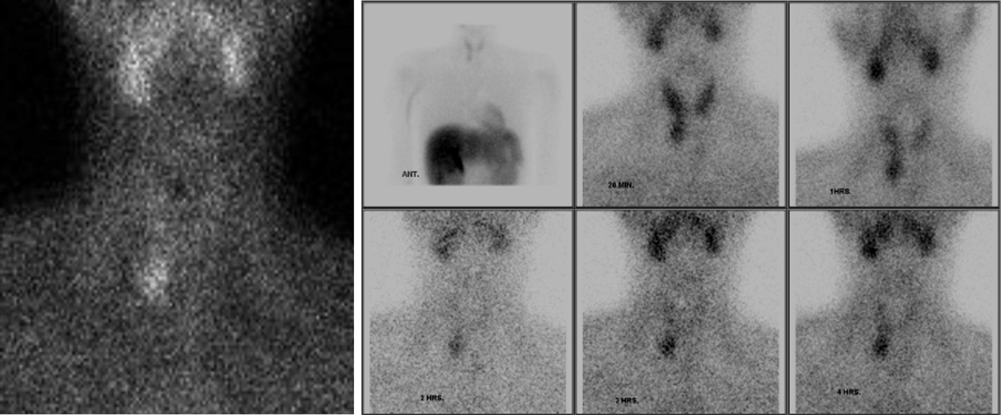
Figure 4 Parathyroid scintigraphy with sestaMIBI.
Radionuclide parathyroid scintigraphy was carried out following the administration of 550 MBq of Tc-99m sestaMIBI by intravenous injection. Multiple static images were taken starting from the time of injection and continuing up to 4 hours later. An area of abnormal tracer deposition is observed in the lower pole of the right thyroid lobe.
- Citation: Karim MM, Raza H, Parkash O. Recurrent acute pancreatitis as an initial presentation of primary hyperparathyroidism: A case report. World J Clin Cases 2024; 12(29): 6302-6306
- URL: https://www.wjgnet.com/2307-8960/full/v12/i29/6302.htm
- DOI: https://dx.doi.org/10.12998/wjcc.v12.i29.6302


