©The Author(s) 2024.
World J Clin Cases. Oct 6, 2024; 12(28): 6132-6136
Published online Oct 6, 2024. doi: 10.12998/wjcc.v12.i28.6132
Published online Oct 6, 2024. doi: 10.12998/wjcc.v12.i28.6132
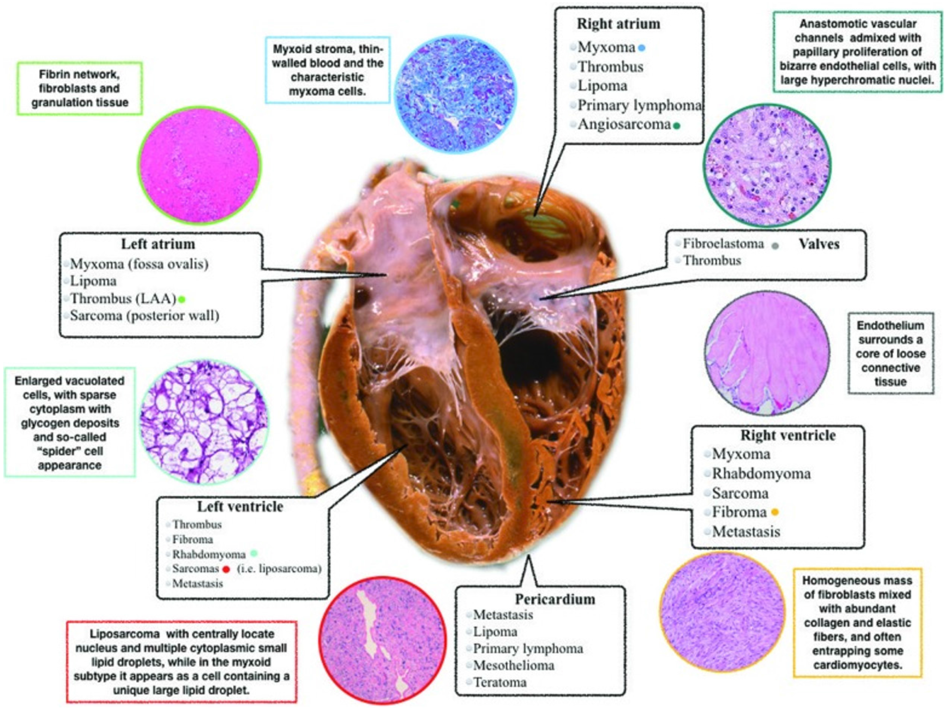
Figure 1 Localization, pathophysiology, and histopathological aspects of cardiac masses.
Citation: Castrichini M, Albani S, Pinamonti B, Sinagra G. Atrial thrombi or cardiac tumours? The image-challenge of intracardiac masses: a case report. Eur Heart J Case Rep 2020; 4: 1-6. Copyright© The Authors 2020. Published by Oxford University Press on behalf of the European Society of Cardiology. The authors have obtained the permission for figure using from the European Heart Journal - Case Reports (Supplementary material).
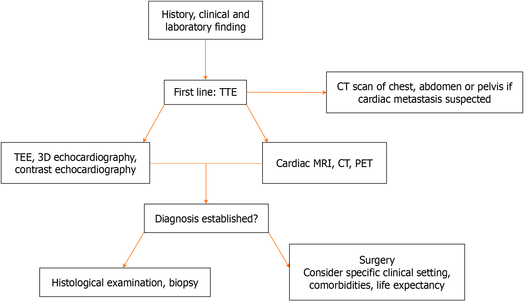
Figure 2 Flow chart of the diagnostic workup in patients with cardiac masses.
CT: Computed tomography; MRI: Magnetic resonance imaging; PET: Positron emission tomography; TEE: Trans esophageal echocardiography; TTE: Trans thoracic echocardiography.
- Citation: De Maria E. Approach to cardiac masses: Thinking inside and outside the box. World J Clin Cases 2024; 12(28): 6132-6136
- URL: https://www.wjgnet.com/2307-8960/full/v12/i28/6132.htm
- DOI: https://dx.doi.org/10.12998/wjcc.v12.i28.6132


