©The Author(s) 2024.
World J Clin Cases. Sep 6, 2024; 12(25): 5769-5774
Published online Sep 6, 2024. doi: 10.12998/wjcc.v12.i25.5769
Published online Sep 6, 2024. doi: 10.12998/wjcc.v12.i25.5769
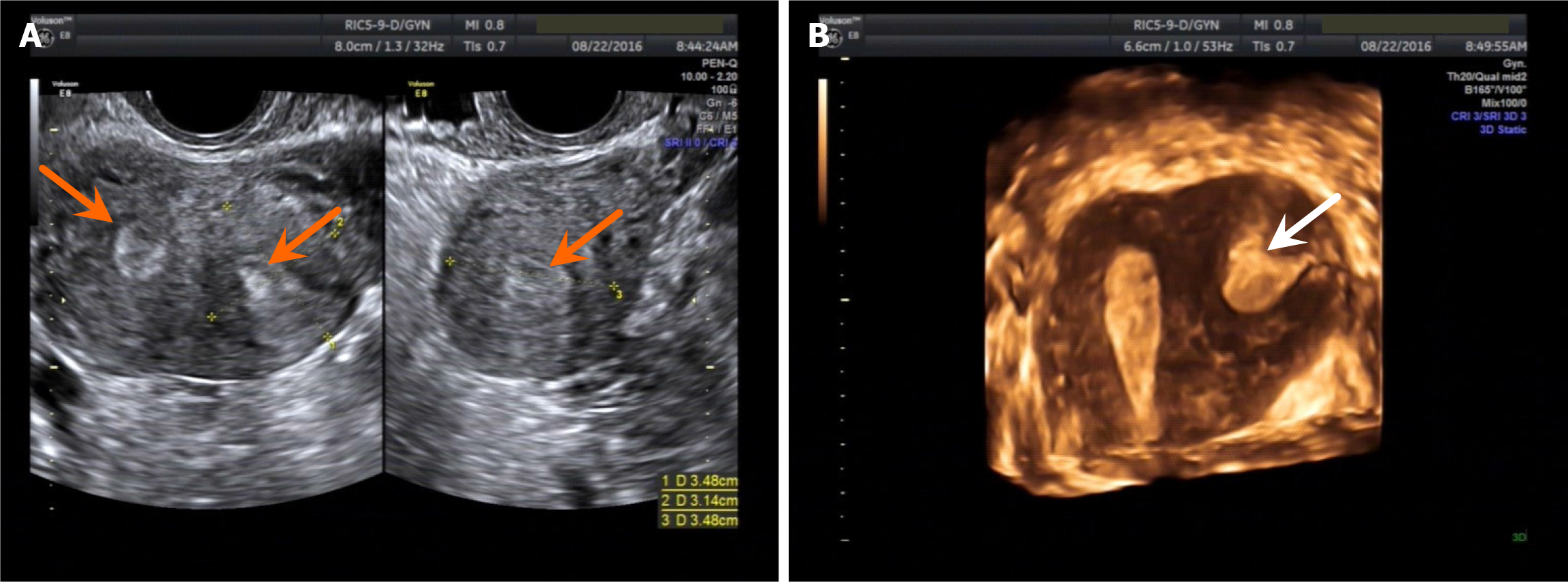
Figure 1 Ultrasound images of Robert’s uterus.
A: Two-dimensional; B: Three-dimensional.
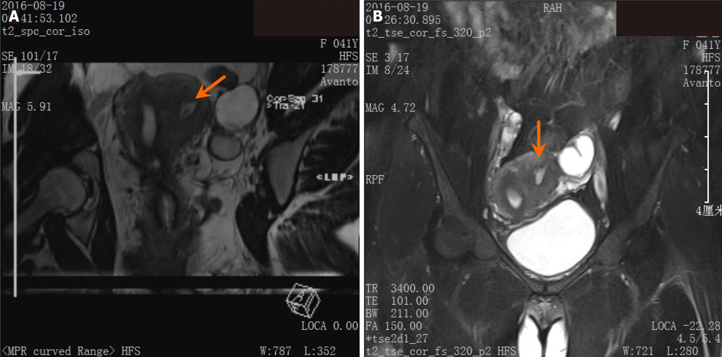
Figure 2 Magnetic resonance imaging of Robert’s uterus.
A: T2 3D reconstruction image showed endometrioid signals and haematosis between the left muscle wall accompanied by adenomyosis of the left uterine wall T2 3D reconstruction image showed endometrioid signals and haematosis between the left muscle wall accompanied by adenomyosis of the left uterine wall; B: Coronal T2-weighted image showed endometrioid signals and haematosis between the left muscle wall, with a peripheral binding zone, accompanied by adenomyosis of the left uterine wall.
Figure 3 Laparoscopy and hysteroscopy.
A: Full protrusion at the left side of the uterus observed during laparoscopy; B: An opening was visible only at the right uterine horn and right fallopian tube during hysteroscopy; C: Lesions in the left blind cavity were removed, and the endometrium in the blind cavity was visible during laparoscopy.
- Citation: Dong J, Wang JJ, Fei JY, Wu LF, Chen YY. Laparoscopy combined with hysteroscopy in the treatment of Robert’s uterus accompanied by adenomyosis: A case report. World J Clin Cases 2024; 12(25): 5769-5774
- URL: https://www.wjgnet.com/2307-8960/full/v12/i25/5769.htm
- DOI: https://dx.doi.org/10.12998/wjcc.v12.i25.5769


