©The Author(s) 2024.
World J Clin Cases. Aug 6, 2024; 12(22): 5245-5252
Published online Aug 6, 2024. doi: 10.12998/wjcc.v12.i22.5245
Published online Aug 6, 2024. doi: 10.12998/wjcc.v12.i22.5245
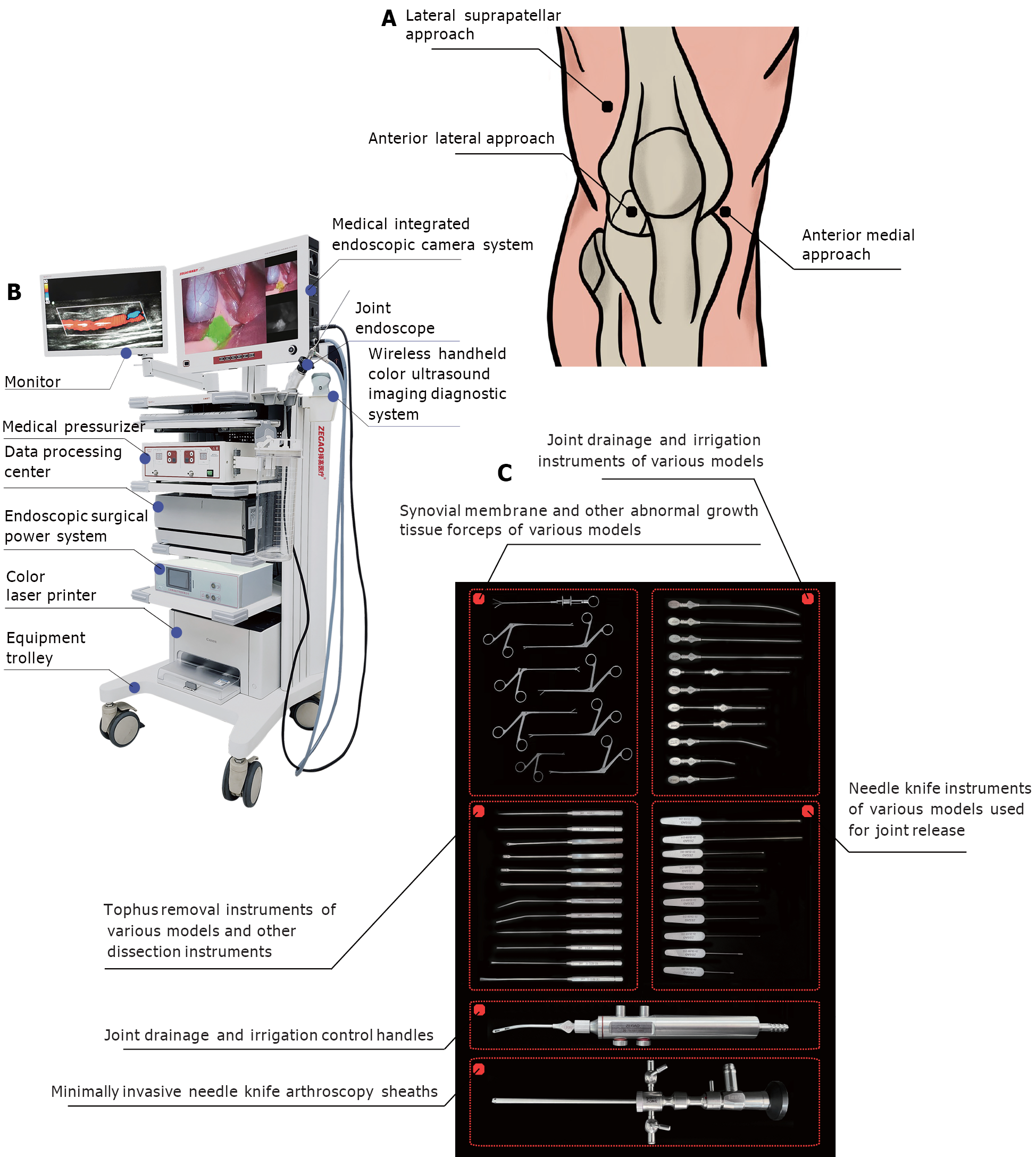
Figure 1 Minimally invasive paths and the minimally invasive needle-knife scope therapy.
A: The surgical pathways; B and C: The surgical instruments.
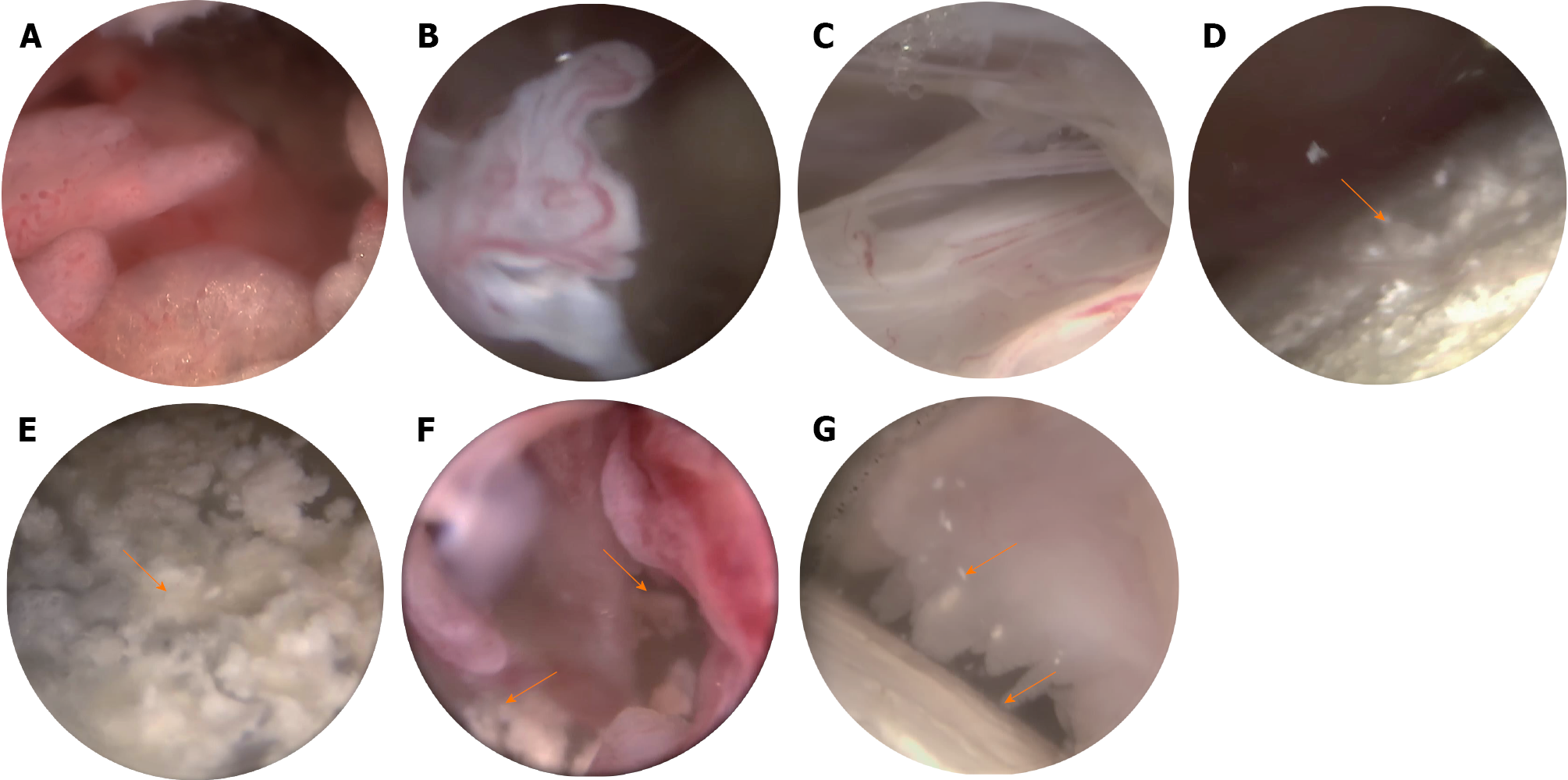
Figure 2 Intra-articular changes in the knee joint.
A-C: Proliferative synovium and pannus; D-G: Intra-articular monosodium urate deposition.
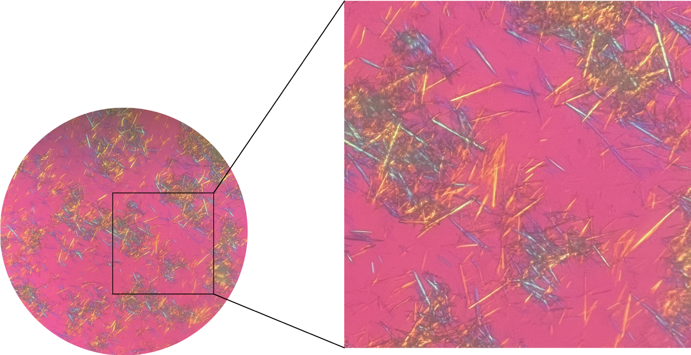
Figure 3 Crystallization diagram of monosodium urate under polarized light microscopy.
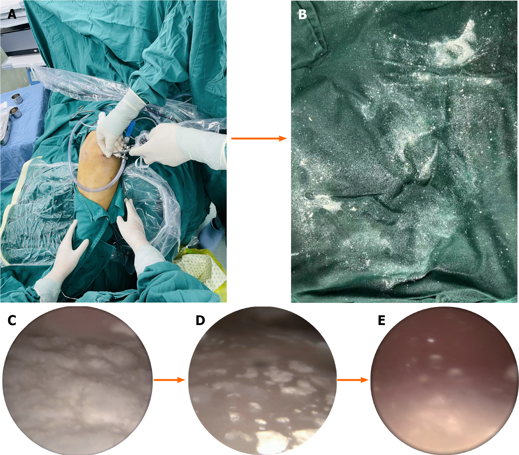
Figure 4 Knee joint debridement process.
A: Surgical process; B: Urate crystals washed out of the body; C: Before clearance; D: During clearance; E: After clearance.
- Citation: Chen ZY, Ou-Yang MH, Li SW, Ou R, Chen ZH, Wei S. Concomitant atypical knee gout and seronegative rheumatoid arthritis: A case report. World J Clin Cases 2024; 12(22): 5245-5252
- URL: https://www.wjgnet.com/2307-8960/full/v12/i22/5245.htm
- DOI: https://dx.doi.org/10.12998/wjcc.v12.i22.5245


