©The Author(s) 2024.
World J Clin Cases. Jul 26, 2024; 12(21): 4762-4769
Published online Jul 26, 2024. doi: 10.12998/wjcc.v12.i21.4762
Published online Jul 26, 2024. doi: 10.12998/wjcc.v12.i21.4762
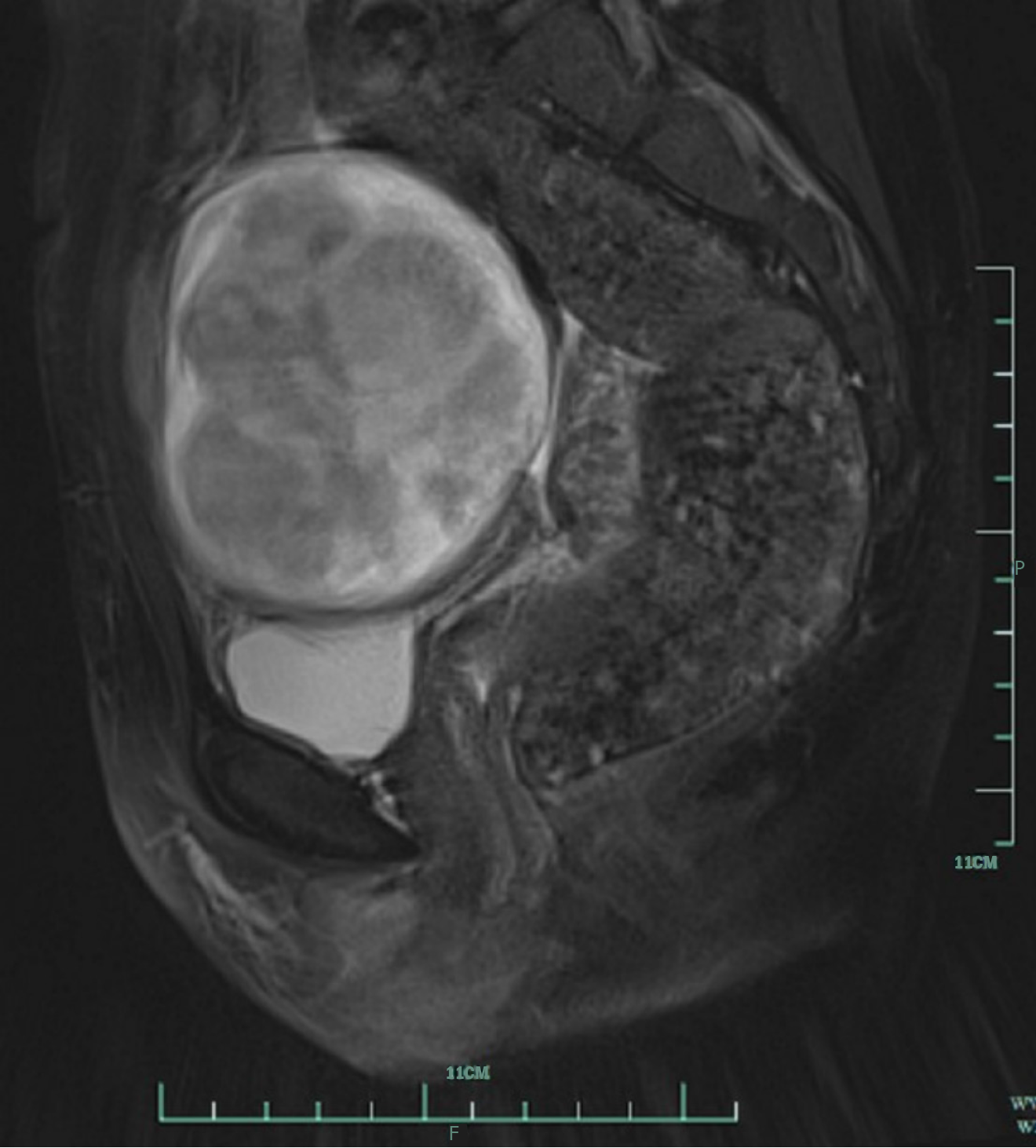
Figure 1
Pelvic magnetic resonance imaging showing an anterior uterine mass, multiple uterine fibroids and slight pelvic effusion.
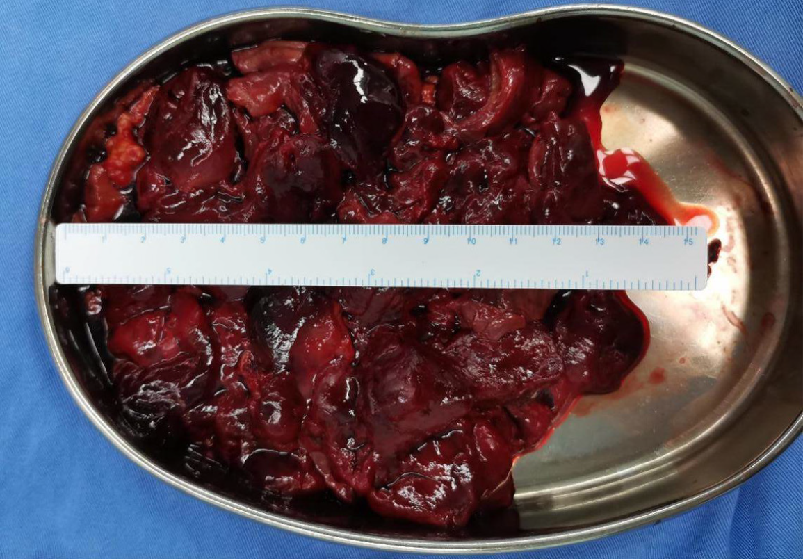
Figure 2
Retroperitoneal leiomyoma removed and morcellated by laparoscopic surgery.
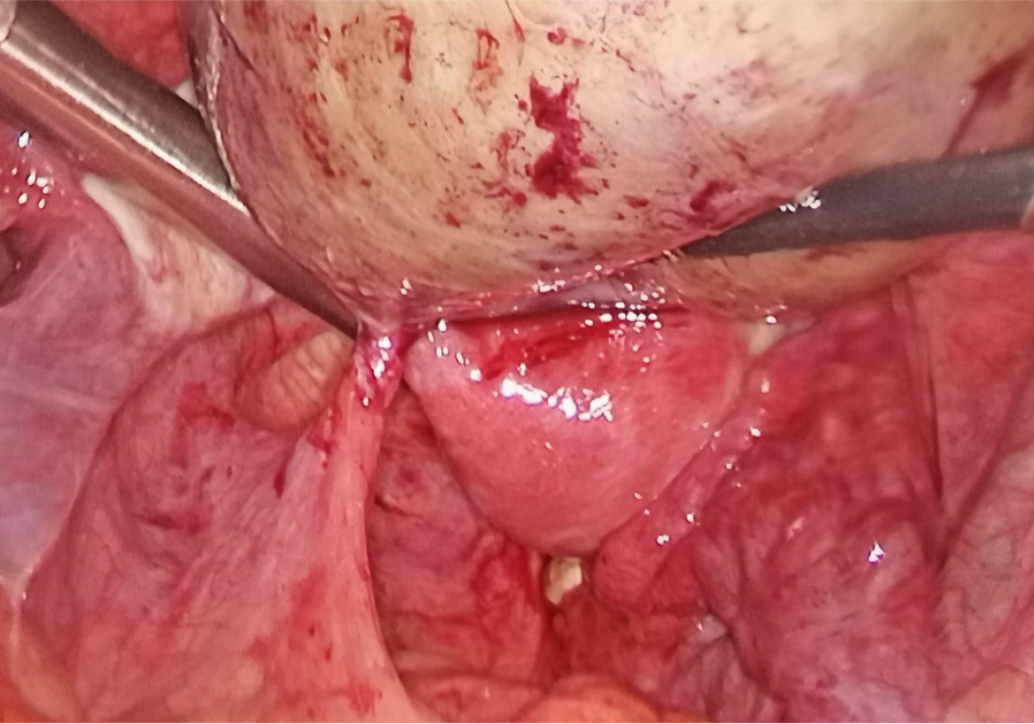
Figure 3
The pedicle of the retroperitoneal leiomyoma was located 2 cm from the posterior peritoneum to the left of the rectum and was torsed two times.
- Citation: Li J, Zhu-Ge YY, Lin KQ. Torsed retroperitoneal leiomyomas: A case report and review of literature. World J Clin Cases 2024; 12(21): 4762-4769
- URL: https://www.wjgnet.com/2307-8960/full/v12/i21/4762.htm
- DOI: https://dx.doi.org/10.12998/wjcc.v12.i21.4762


