©The Author(s) 2024.
World J Clin Cases. Jun 6, 2024; 12(16): 2856-2861
Published online Jun 6, 2024. doi: 10.12998/wjcc.v12.i16.2856
Published online Jun 6, 2024. doi: 10.12998/wjcc.v12.i16.2856
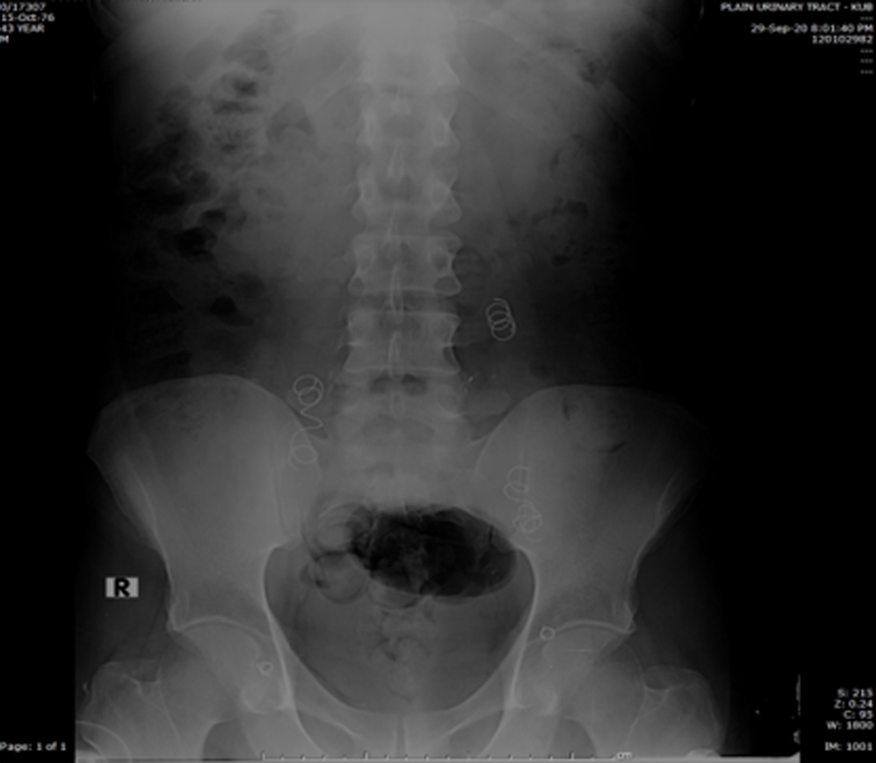
Figure 1 Plain abdominal X-ray (kidneys, ureters and urinary bladder) showing refilling of the coils bilaterally for previous surgery of varicocele.
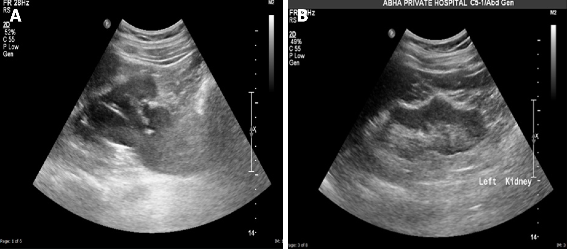
Figure 2 Ultrasonography of the left kidney showing hydronephrosis and ultrasonography after surgical repair.
A: Ultrasound of the left kidney showing hydronephrosis; B: Ultrasound after surgical repair (end-to-end ureteric anastomosis) showing normal kidney with no residual hydronephrosis.
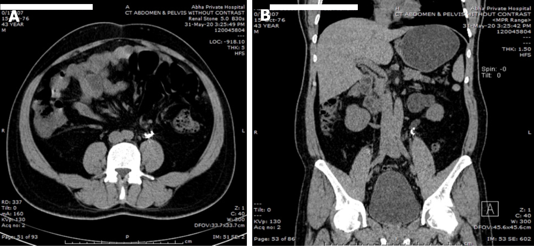
Figure 3 Computed tomography of the abdomen.
A: Axial; B: Coronal views showing the coils and left hydro ureter.
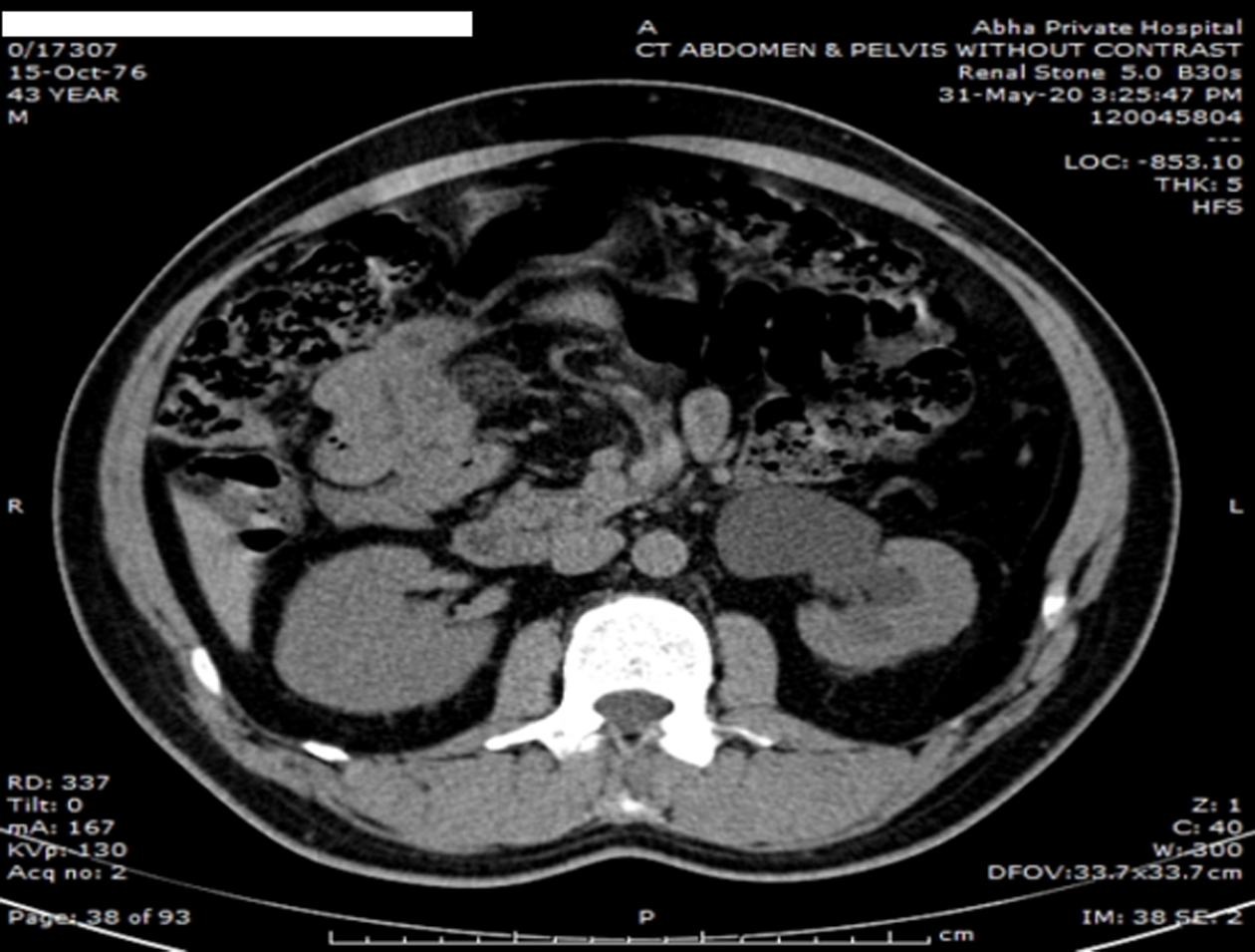
Figure 4 Computed tomography scan axial view showing left hydronephrosis due to obstructed ureter by the varicocele coils.
Figure 5 Intra operative photos showing the coils.
A: Intra operative laparoscopic view showing migration and erosion of the coils through gonadal vein wall; B: Another laparoscopic view showing grasping and removal of the coils; C: Double J stent inside the ureter during the end-to-end ureteric anastomosis.
- Citation: Alamri A. Migration of varicocele coil leading to ureteral obstruction and hydronephrosis: A case report. World J Clin Cases 2024; 12(16): 2856-2861
- URL: https://www.wjgnet.com/2307-8960/full/v12/i16/2856.htm
- DOI: https://dx.doi.org/10.12998/wjcc.v12.i16.2856


