©The Author(s) 2024.
World J Clin Cases. May 26, 2024; 12(15): 2642-2648
Published online May 26, 2024. doi: 10.12998/wjcc.v12.i15.2642
Published online May 26, 2024. doi: 10.12998/wjcc.v12.i15.2642
Figure 1 Computed tomography scan.
A: Computed tomography (CT) scan of the head. Calcified nodules located beneath the ependymal membrane of both lateral ventricles; symmetrically distributed (orange arrow); B: Abdominal CT enhancement. Multiple regular and well-defined nodules in both kidneys, with visible fat density inside. After enhancement, the solid components are enhanced, but the fat components are not enhanced (orange arrow); C: Chest CT scan. An irregular ground glass opacity nodule in the posterior basal segment of the right lower lobe of the lung (orange arrow).
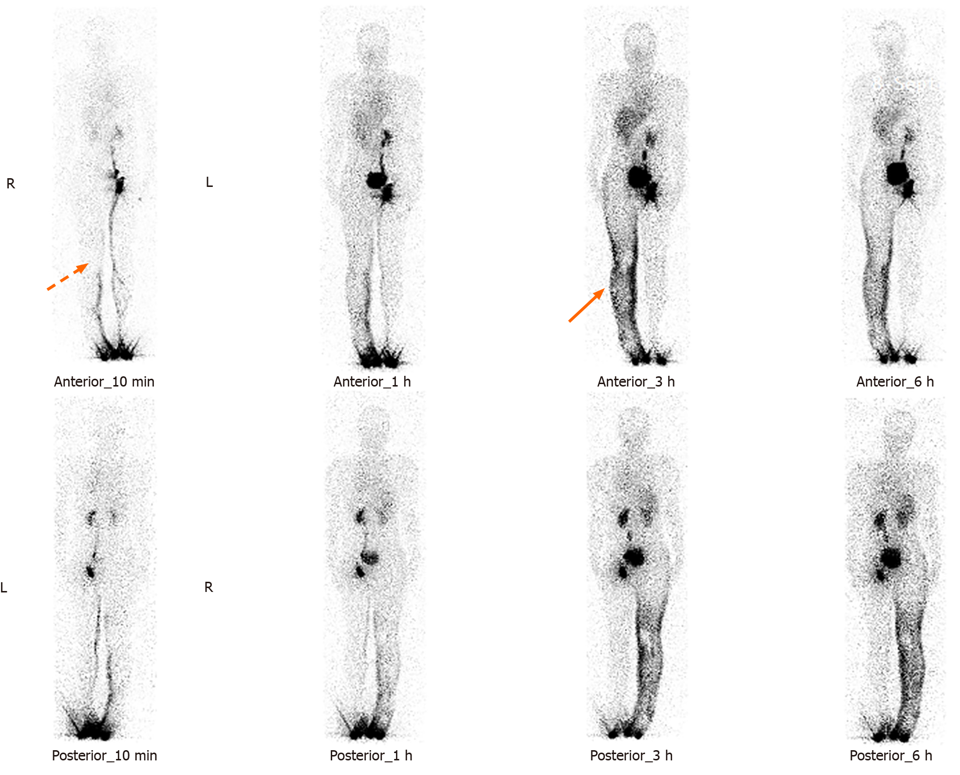
Figure 2 99Tcm-DX-lymphoscintigraphy: The imaging agent was diffusely and unevenly distributed in the right lower limb, waist, buttocks, and external genital area (orange arrow).
The lymphatic vessels in the right lower limb were not visualized, and the right inguinal, iliac, and lumbar lymphatic vessels and lymph nodes were not visualized (orange dashed arrow).
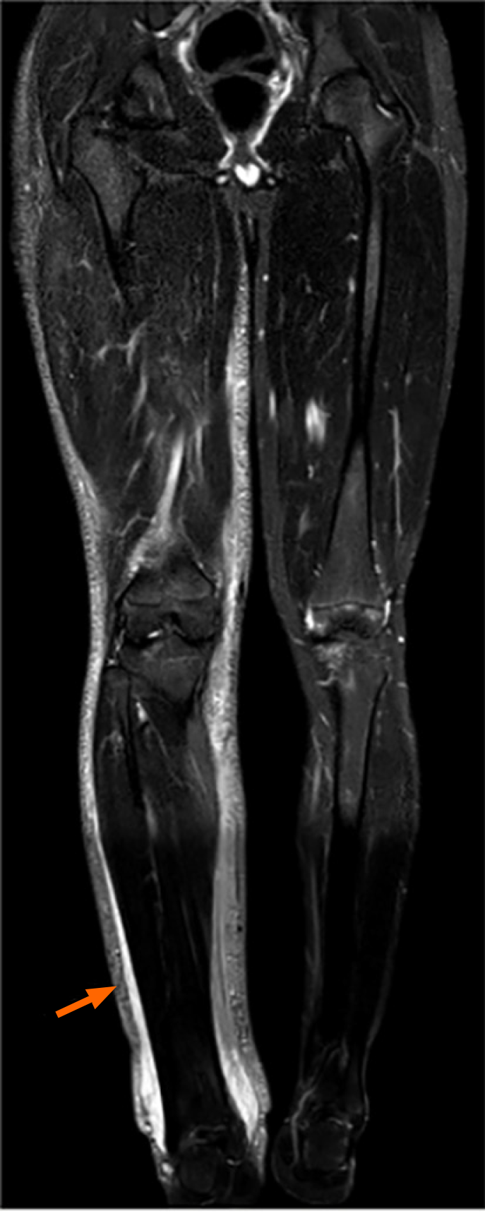
Figure 3 Coronal view of the short time inversion recovery sequence of the entire lower limb.
Compared with those on the left, the skin on the right lower limb was thickened, and the subcutaneous soft tissue was swollen, with abnormally high signal shadows visible inside (orange arrows).
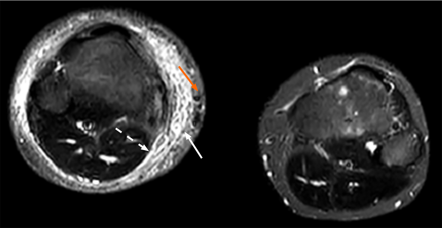
Figure 4 Short time inversion recovery sequence transverse section of lower limb magnetic resonance imaging: Signs of lymphedema in subcutaneous soft tissue, including the honeycomb sign (white arrow) and band sign (white dashed arrow).
Thickened, tortuous and dilated veins were observed (orange arrow).
- Citation: Li XP, Sun XL, Liu X, Wen Z, Jiang LH, Fu Y, Yue YL, Wang RG. Tuberous sclerosis complex combined with primary lymphedema: A case report. World J Clin Cases 2024; 12(15): 2642-2648
- URL: https://www.wjgnet.com/2307-8960/full/v12/i15/2642.htm
- DOI: https://dx.doi.org/10.12998/wjcc.v12.i15.2642


