Copyright
©The Author(s) 2024.
World J Clin Cases. May 26, 2024; 12(15): 2597-2605
Published online May 26, 2024. doi: 10.12998/wjcc.v12.i15.2597
Published online May 26, 2024. doi: 10.12998/wjcc.v12.i15.2597
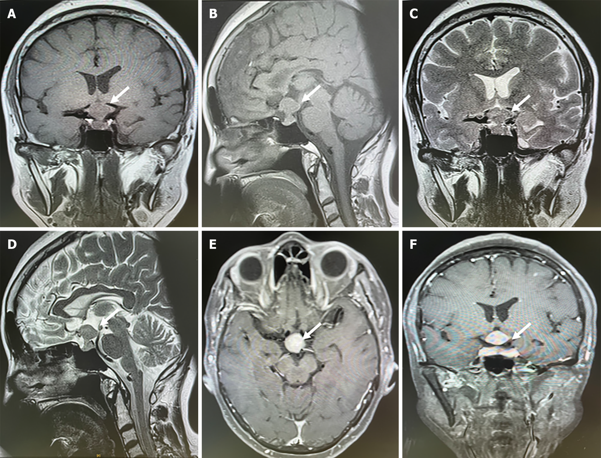
Figure 1 Sellar magnetic resonance imaging showed a sellar lesion related to carotid artery.
A-D: The leision located in the sellar region presented with an isointense signal on T1 and T2-weighted images and could see hourglass sign; E and F: The leision was intensified homogenously enhanced after contrast magnetic resonance imaging.
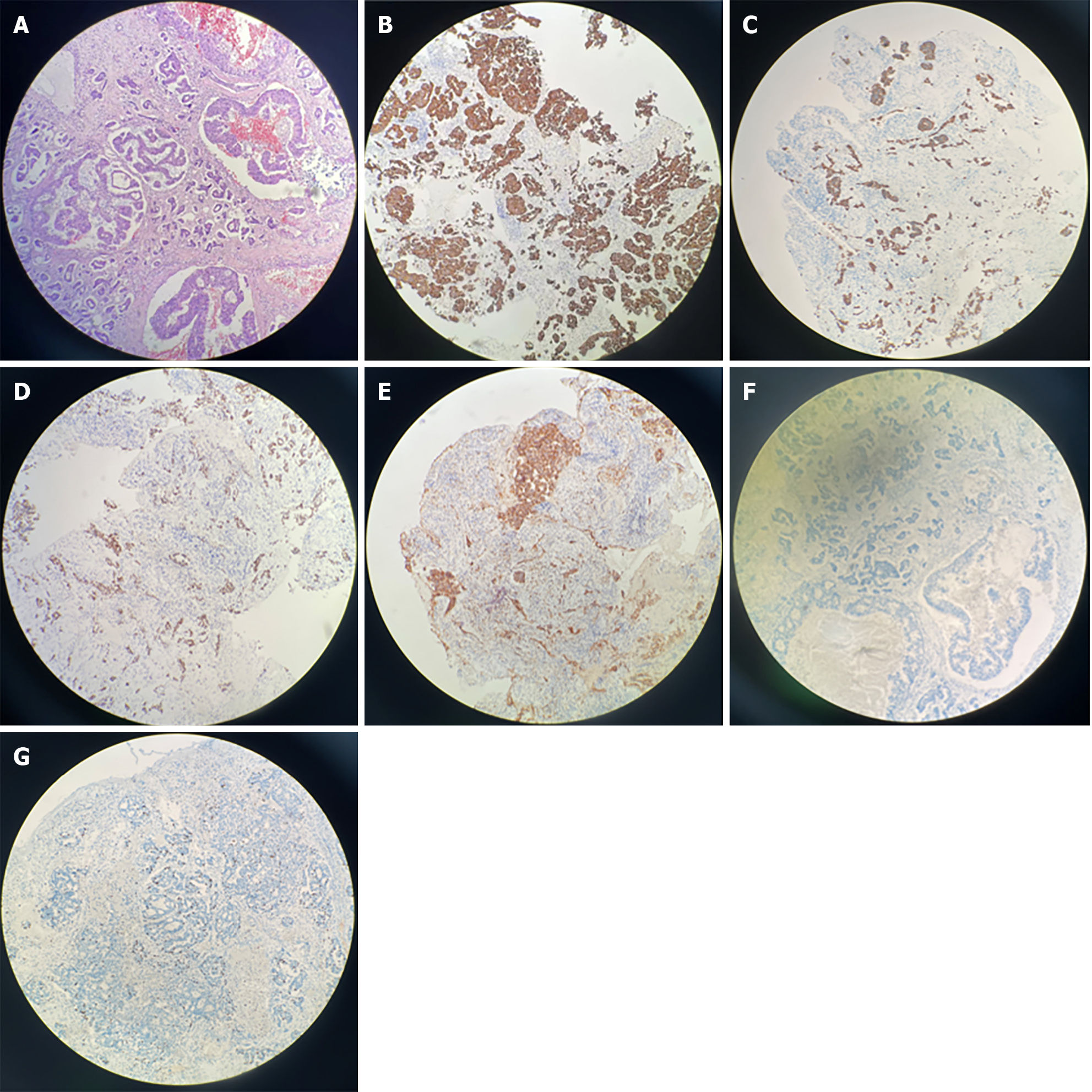
Figure 2 The histological features of the lesions revealed pituitary metastasis from lung adenocarcinoma.
A: Hematoxylin and eosin staining found epithelial neoplasm and considered metastatic adenocarcinoma (× 100); B-F: Immunohistochemistry revealed positive for CK7, CK18, TTF-1, NapsinA and GATA3 (× 100); G: Immunohistochemical staining of Ki-67 (× 100).
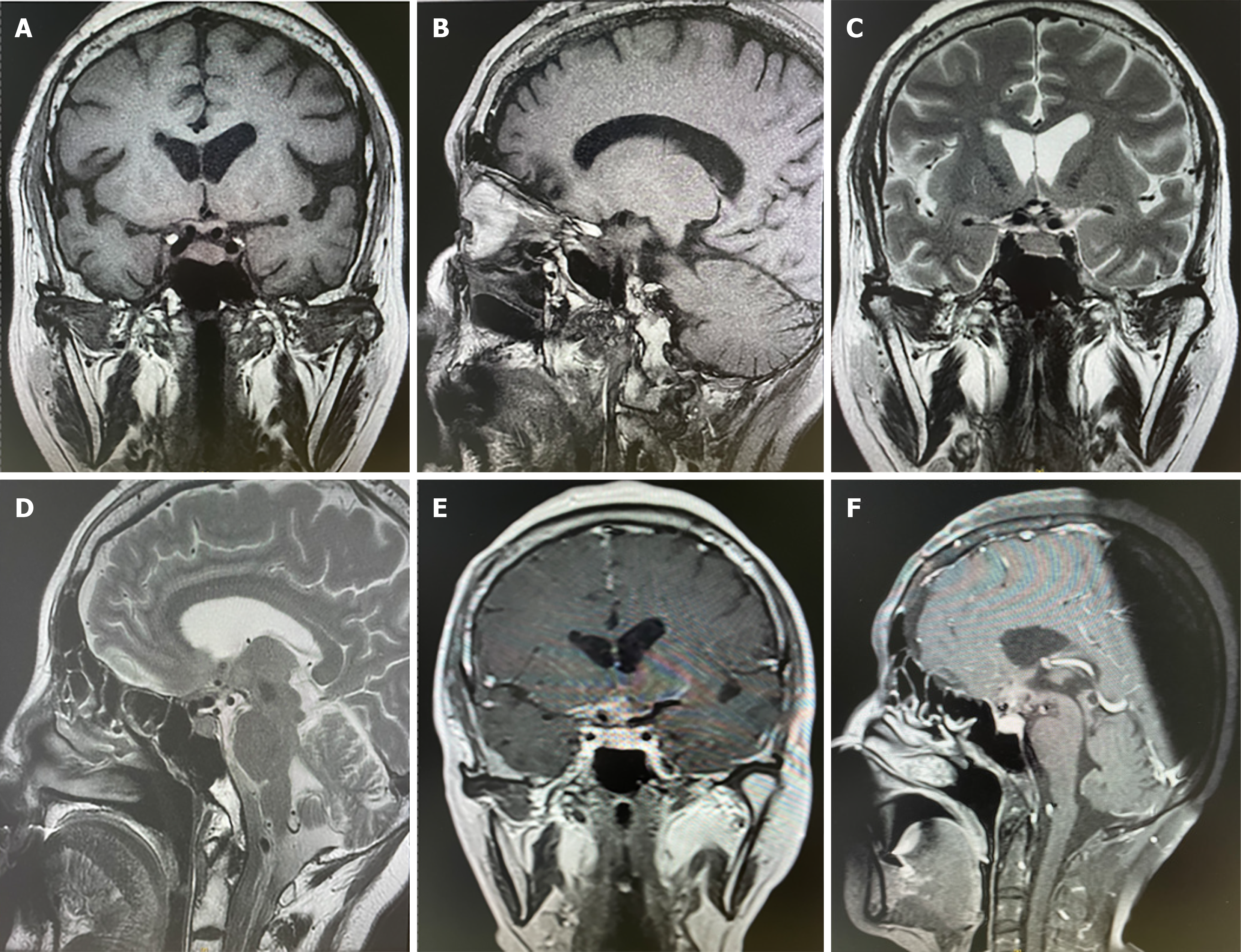
Figure 3 Sellar magnetic resonance imaging showed no neoplasm recurrence in 3 months after surgery.
A-D: Postoperative changes of sellar region and there was no leision found in T1 and T2-weighted images; E and F: There was no intensified leision imaging after contrast magnetic resonance imaging.
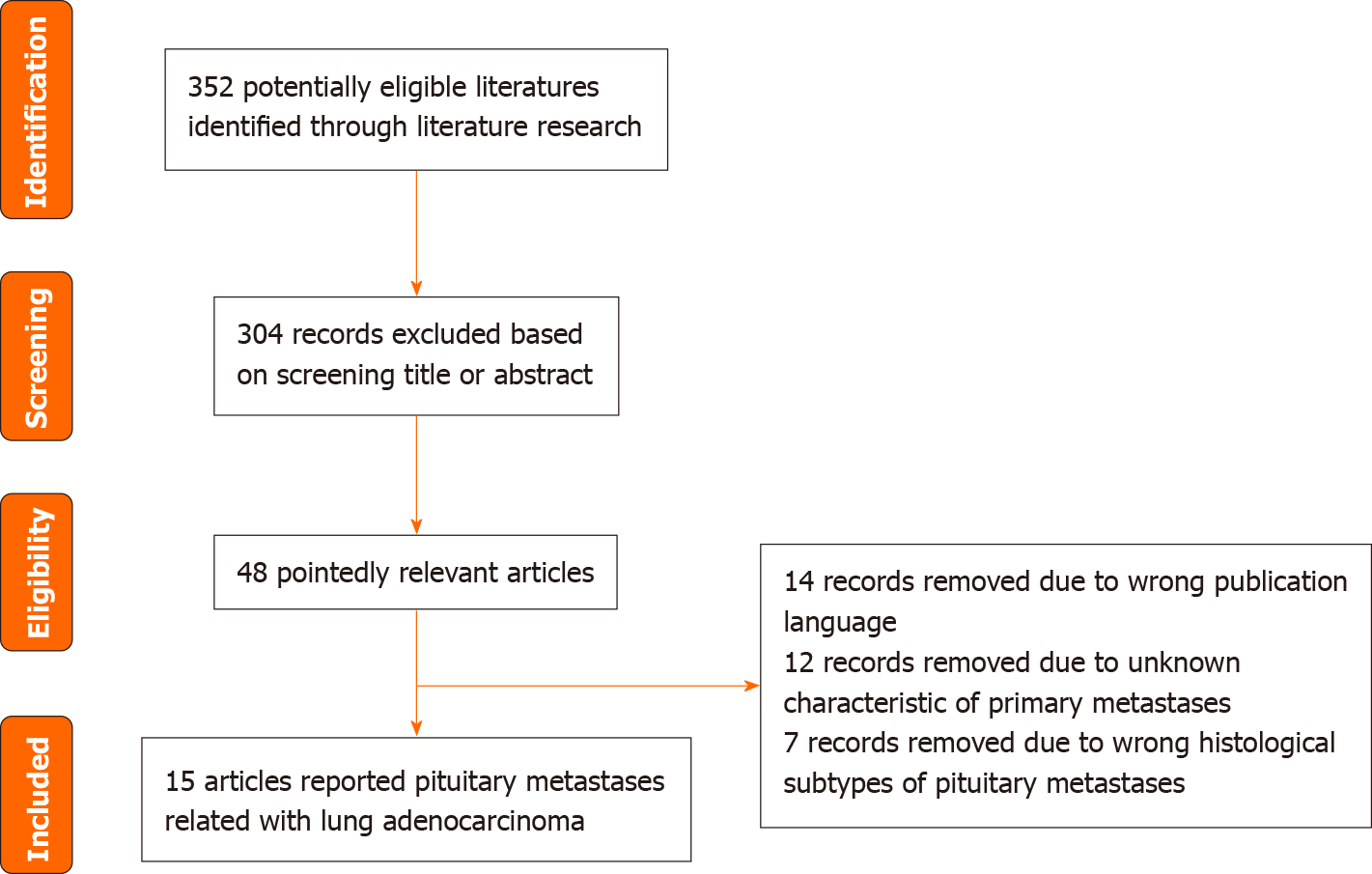
Figure 4 Flowchart of the study selection.
- Citation: Wang Q, Liu XW, Chen KY. Pituitary metastasis from lung adenocarcinoma: A case report. World J Clin Cases 2024; 12(15): 2597-2605
- URL: https://www.wjgnet.com/2307-8960/full/v12/i15/2597.htm
- DOI: https://dx.doi.org/10.12998/wjcc.v12.i15.2597


