©The Author(s) 2024.
World J Clin Cases. May 26, 2024; 12(15): 2586-2596
Published online May 26, 2024. doi: 10.12998/wjcc.v12.i15.2586
Published online May 26, 2024. doi: 10.12998/wjcc.v12.i15.2586
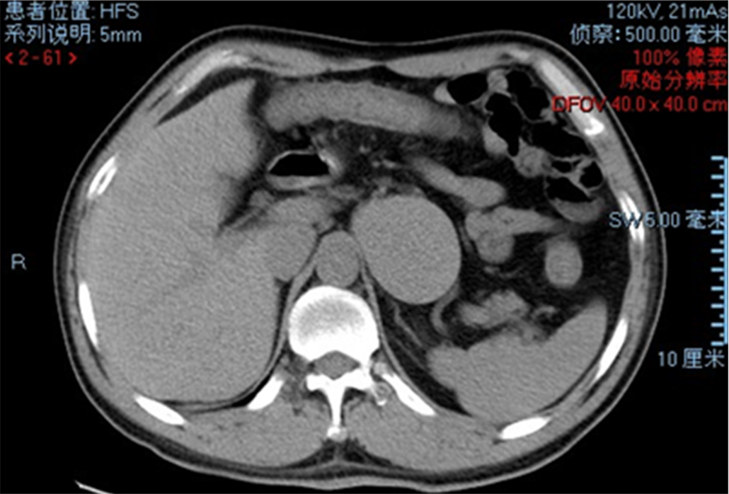
Figure 1 Abdominal noncontrast computed tomography scan depicting a sizable cystic formation.
The scan also revealed scattered calcifications throughout the abdominal and pelvic regions.
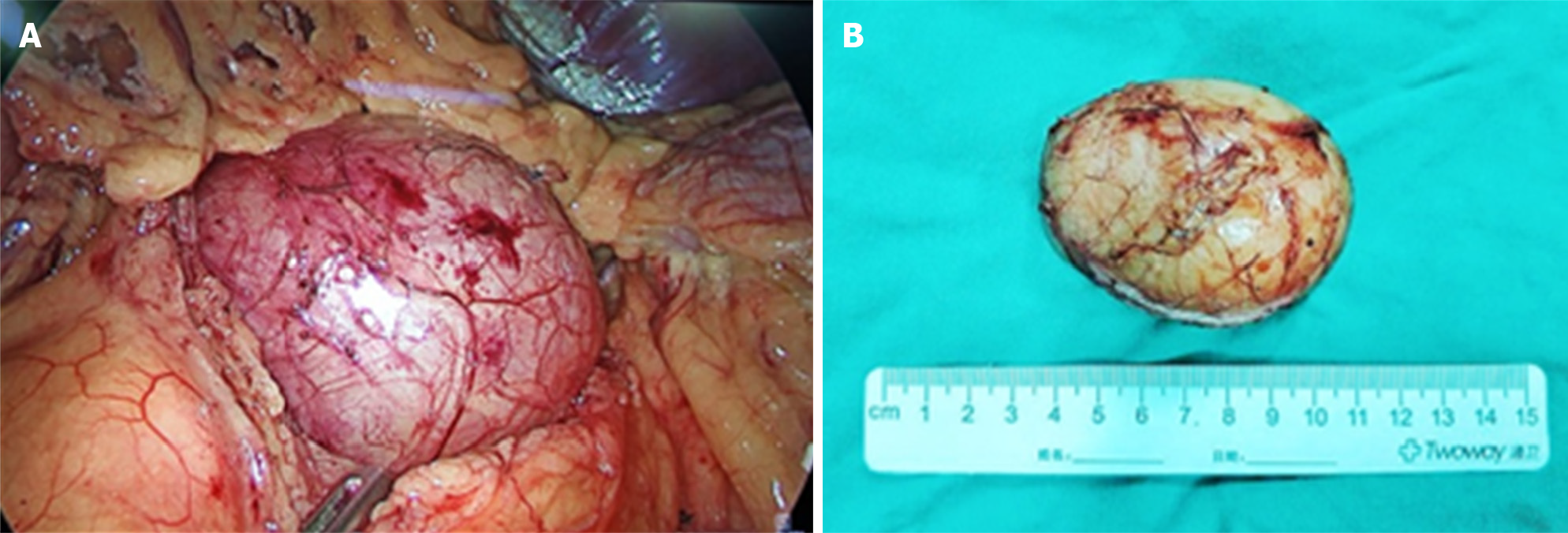
Figure 2 Cystic-solid tumor.
A: Relationship of cystic lesions with surrounding structures and their maximum diameter; B: Complete surgical removal of the cystic-solid tumor, measuring 8.8 cm in length, 7.2 cm in width, and 2.6 cm in depth.
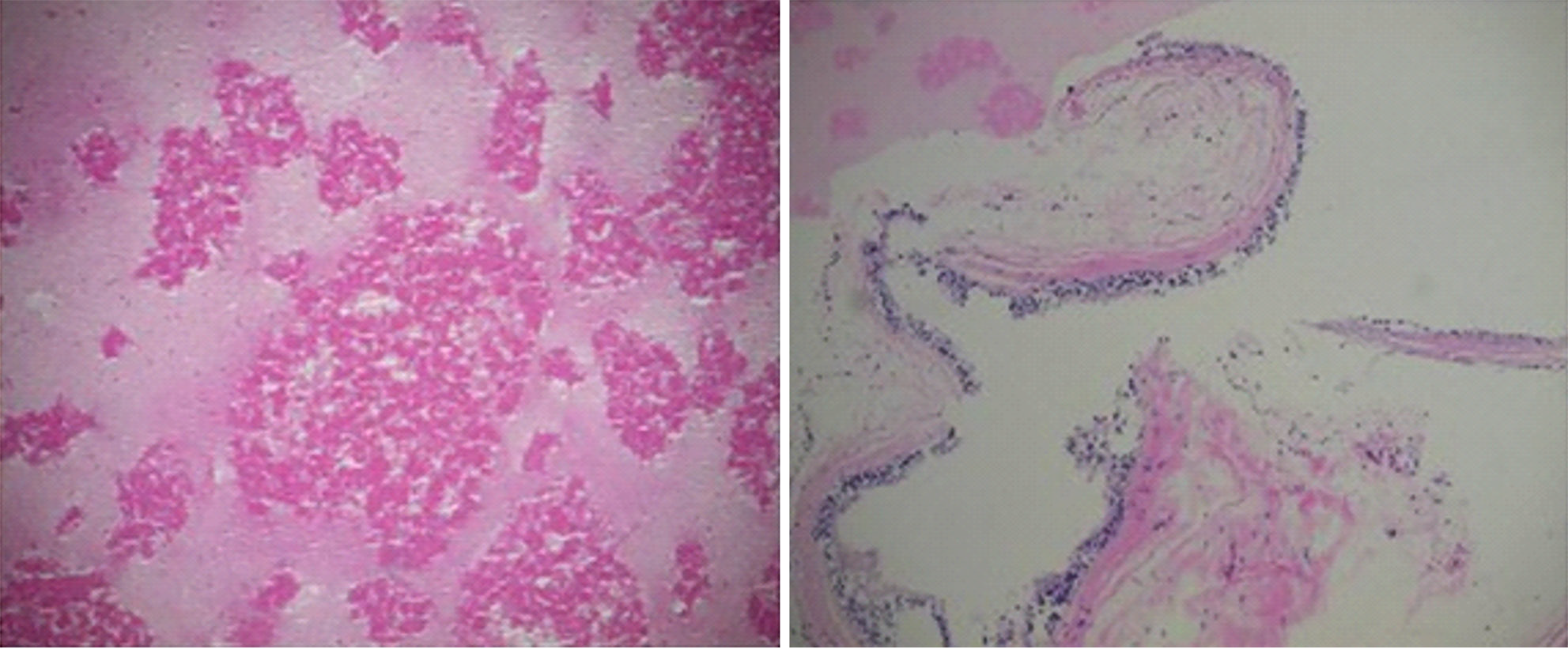
Figure 3 Histopathological analysis showing a retroperitoneal mass identified as an ectopic bronchogenic cyst, featuring nodular tissue in shades of gray-white and yellow.
The cystic portion contains a light yellow, soft, gelatinous substance, encased by a smooth, encapsulated surface.
- Citation: Malik A, Naseer QA, Iqbal MA, Han SY, Dang SC. Retroperitoneal bronchogenic cyst: A case report and review of literature. World J Clin Cases 2024; 12(15): 2586-2596
- URL: https://www.wjgnet.com/2307-8960/full/v12/i15/2586.htm
- DOI: https://dx.doi.org/10.12998/wjcc.v12.i15.2586


