©The Author(s) 2024.
World J Clin Cases. May 26, 2024; 12(15): 2466-2474
Published online May 26, 2024. doi: 10.12998/wjcc.v12.i15.2466
Published online May 26, 2024. doi: 10.12998/wjcc.v12.i15.2466
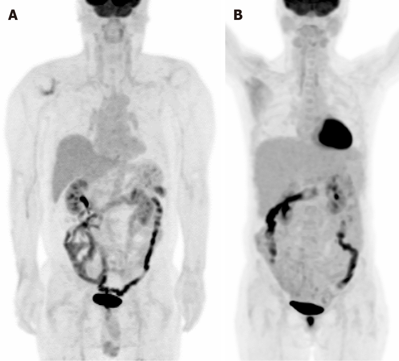
Figure 1 Diffuse physiological bowel fluorine-18 fluorodeoxyglucose uptake.
Images depicting fluorine-18 fluorodeoxyglucose uptake in the bowel. A: Diffuse intestinal uptake observed in a 58-year-old male patient diagnosed with B-cell lymphoblastic lymphoma; B: Regions of hypermetabolism are highlighted in the hepatic flexure area and descending colon of a 51-year-old woman with a history of gastric cancer. Colonoscopy examination yielded no evidence of abnormalities such as tumors or inflammation, confirming the physiological nature of the observed bowel uptake.
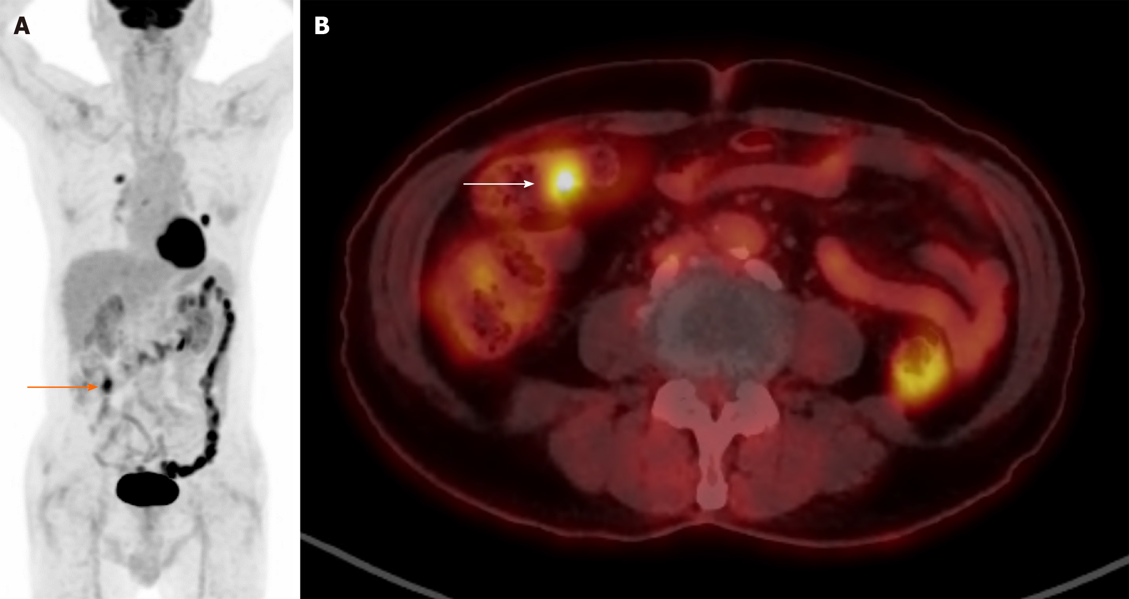
Figure 2 Focal and diffuse intestinal fluorine-18 fluorodeoxyglucose uptake.
A 87-year-old man diagnosed with non-small cell lung cancer (squamous cell carcinoma) is depicted. A: Focal (arrow) and diffuse intestinal uptakes are evident. While diffuse uptake may represent physiological activity, a focal hypermetabolic area raises suspicion for a potential pathological lesion; B: Focal uptake is specifically noted in the proximal transverse colon. Subsequent colonoscopy was performed, revealing no significant abnormal lesions.
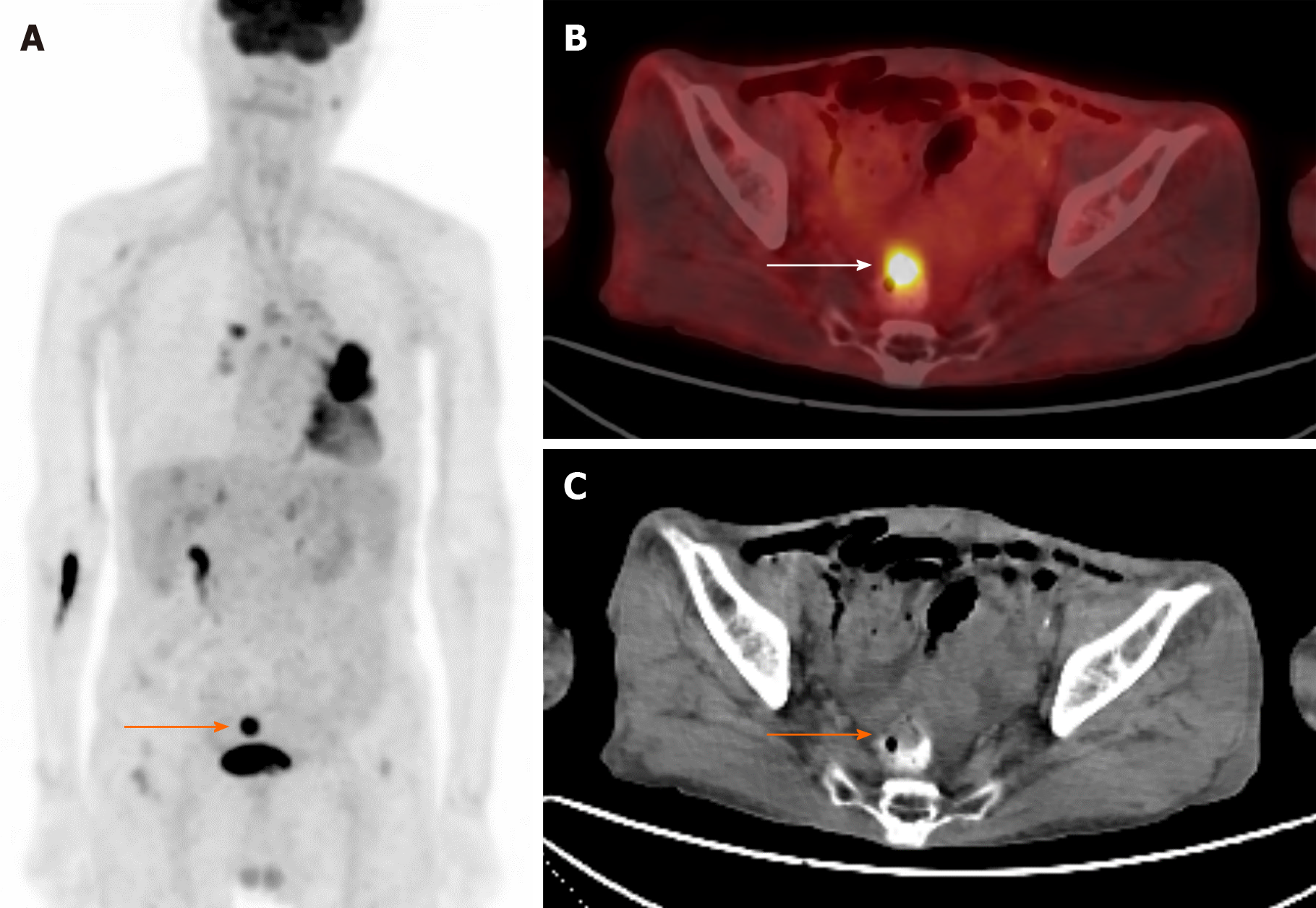
Figure 3 Focal intestinal fluorine-18 fluorodeoxyglucose uptake.
A: Maximum intensity projection depicting focal hypermetabolism superior to the urinary bladder (arrow) in an 88-year-old man diagnosed with non-small cell lung cancer; B: Axial view revealing focal hypermetabolism localized in the rectum; C: Non-contrast-enhanced computed tomography obtained from positron emission tomography/computed tomography, highlighting a space-occupying lesion within the rectum. Notably, a discernible posterior radioopaque area suggests the presence of residual contrast material from a prior contrast-enhanced examination. Subsequent colonoscopy and pathological analysis definitively identified the lesion as adenocarcinoma originating from the rectum.
- Citation: Lee H, Hwang KH. Focal incidental colorectal fluorodeoxyglucose uptake: Should it be spotlighted? World J Clin Cases 2024; 12(15): 2466-2474
- URL: https://www.wjgnet.com/2307-8960/full/v12/i15/2466.htm
- DOI: https://dx.doi.org/10.12998/wjcc.v12.i15.2466


