Copyright
©The Author(s) 2024.
World J Clin Cases. May 16, 2024; 12(14): 2431-2437
Published online May 16, 2024. doi: 10.12998/wjcc.v12.i14.2431
Published online May 16, 2024. doi: 10.12998/wjcc.v12.i14.2431
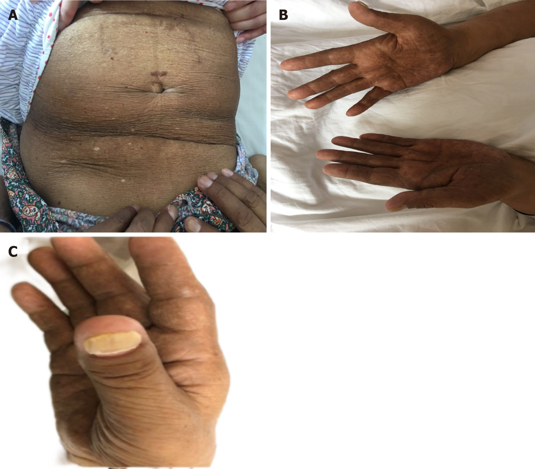
Figure 1 Before initial treatment.
A and B: Diffuse melanosis of the skin; C: Thickened, brittle, and yellow nails.
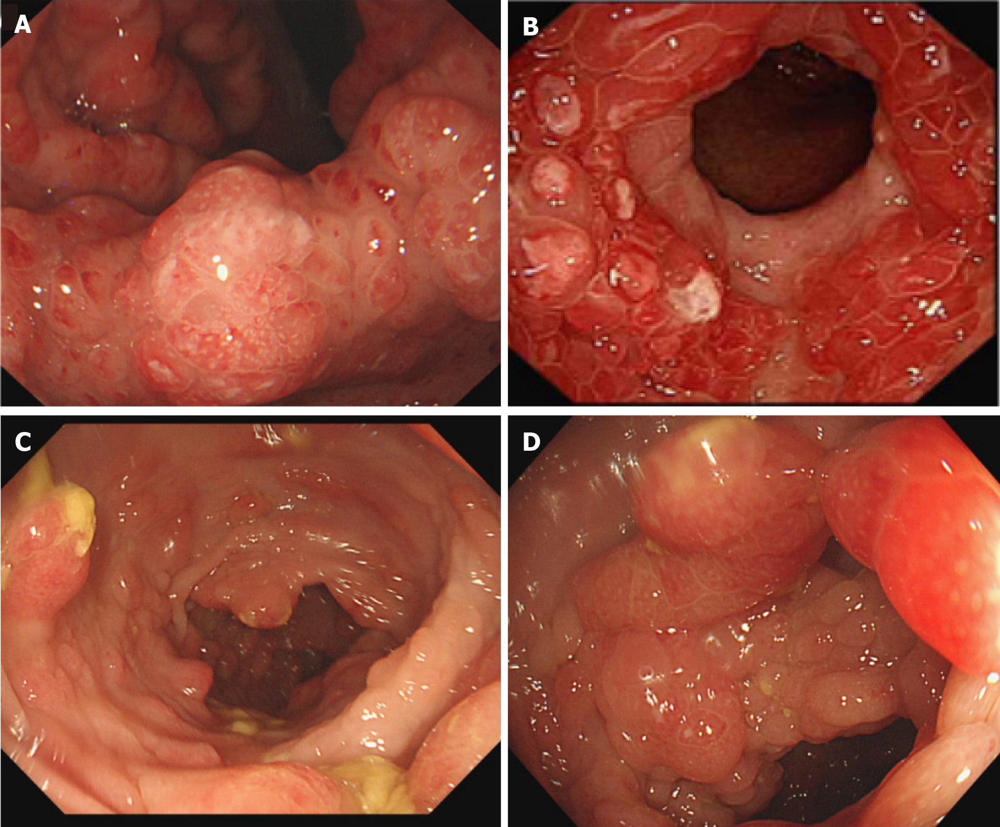
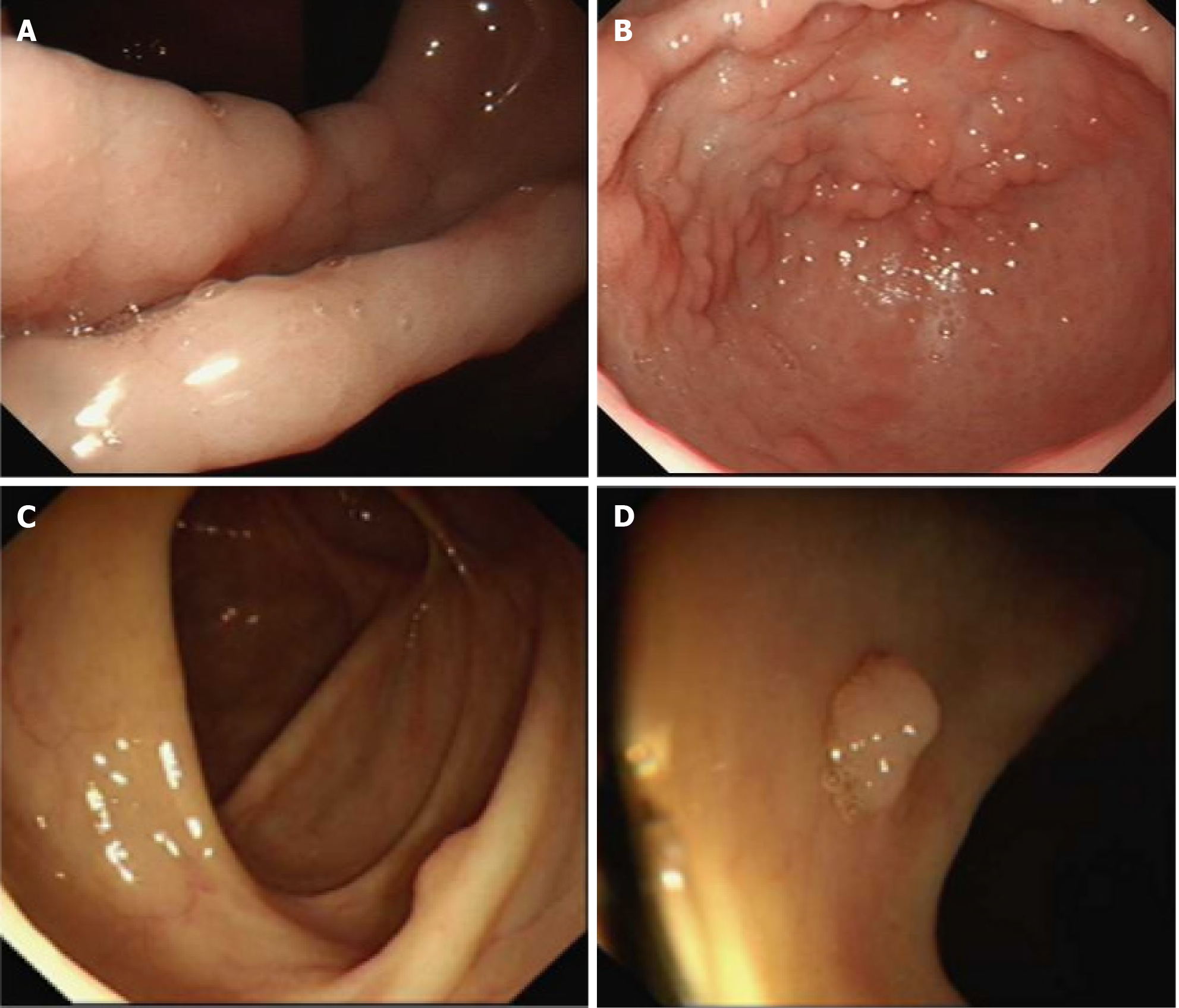
Figure 2 Endoscopic images before treatment.
A and B: The initial gastrointestinal endoscopy showed multiple reddish inflammatory polyps and edematous adjacent mucosa in the angle and antrum of the stomach; C and D: Preliminary colonoscopy showed multiple red inflammatory polyps and adjacent mucosal edema in the colon and rectum.
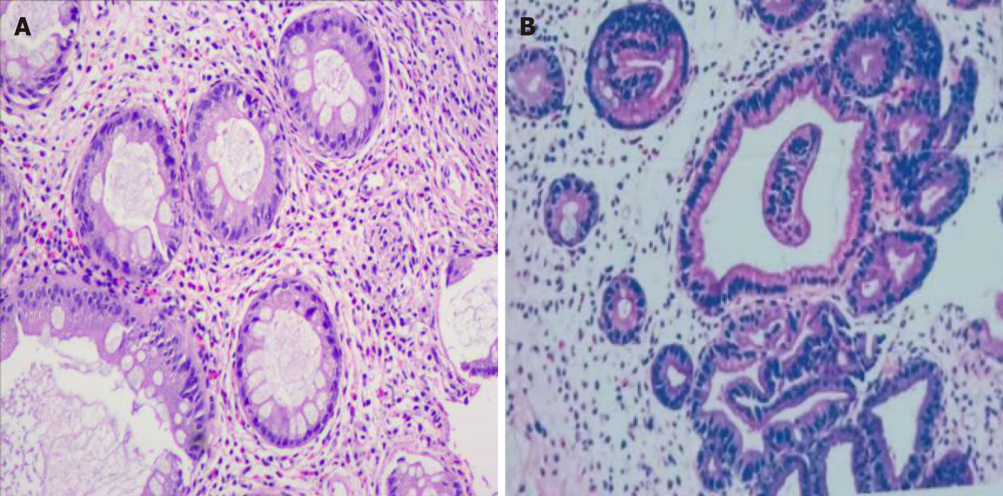
Figure 3
Polyps revealed prominent cystic dilation of the crypts and expanded inflamed lamina propria, consistent with inflammatory polyps.
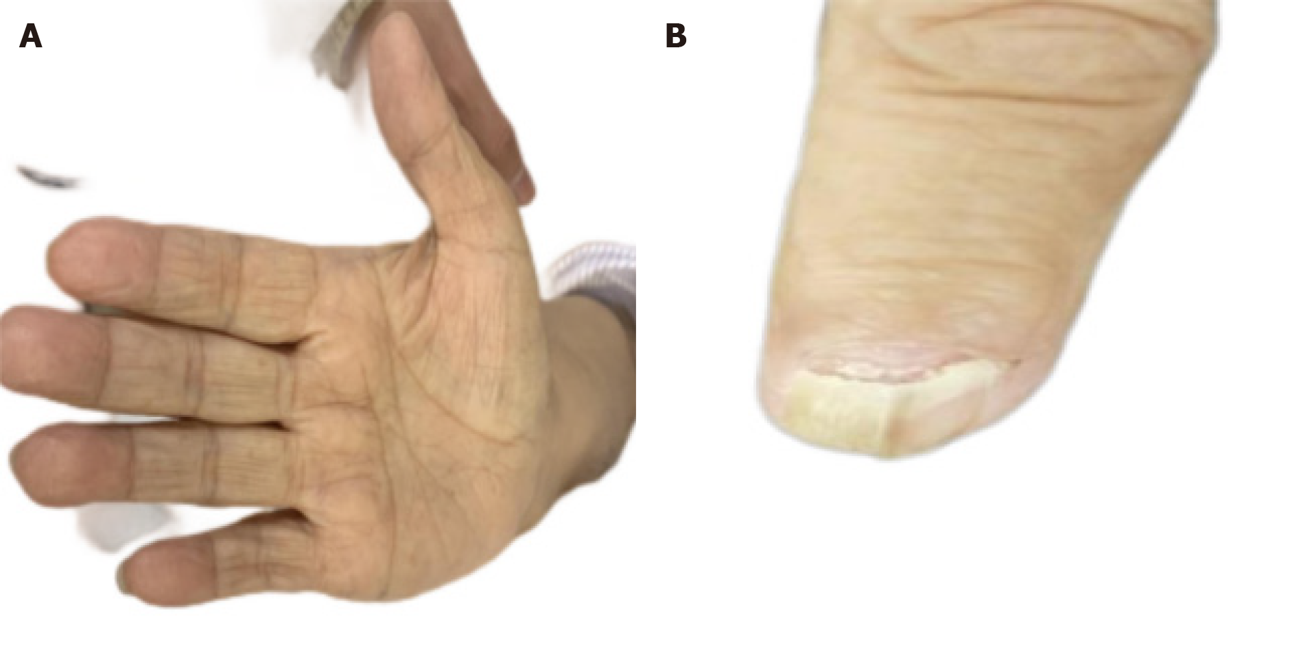
Figure 4 After 3 mo of initial treatment.
A: Normal skin color; B: No improvement in the nails.
Figure 5 Endoscopic images after treatment.
A and B: Three months after treatment, there was a marked improvement in polyp number in the angle and antrum of the stomach; C and D: The intestinal cavity displayed scattered polyps.
- Citation: Chen YL, Wang RY, Mei L, Duan R. Sustained remission of Cronkhite-Canada syndrome after corticosteroid and mesalazine treatment: A case report. World J Clin Cases 2024; 12(14): 2431-2437
- URL: https://www.wjgnet.com/2307-8960/full/v12/i14/2431.htm
- DOI: https://dx.doi.org/10.12998/wjcc.v12.i14.2431


